Classification[edit | edit code]
Myiases are divided into random
,
facultative
and
obligate
.
Based on the location of parasitism, they are divided into tissue
and
cavitary
.
Random
Myiasis is caused by the larvae of those species of flies that normally develop in rotting substances.
They enter the human body accidentally. Flies lay eggs on food, linen, etc. The larvae that emerge from the eggs can be swallowed along with food. Larvae can crawl from contaminated laundry into the urethra. As a result, random cavitary myiases ( intestinal
and
genitourinary
) develop.
Intestinal myiasis[edit | edit code]
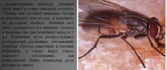
With low acidity and aerophagy, larvae swallowed with food develop in the human intestine. However, more often they die after a few days and are excreted through vomiting or excrement. Intestinal myiases are caused by the larvae of many species of flies, including the housefly ( Musca domestica
), housefly (
Muscina stabulans
), small housefly (
Fannia canicularis
), blue blowfly (
Calliphora vicina
), green blowfly (
Lucilla sericata
), cheese fly (
Piophila casei
), fruit flies (family Drosophilidae), etc. .
Severe intestinal myiases are caused by cheese fly and Drosophila larvae.
With intestinal myiases, the disease is usually acute, but with repeated infections it can take a protracted course. In the United States, such infestations are most often caused by flies of the genus Tubifera tenax. Invasion of larvae into the intestinal mucosa can also develop when infected with flies of the genus Sarcophaga.
There is irritation of the intestinal mucous membranes and their inflammation, accompanied by pain in the abdomen, sometimes in the anus, tenesmus, diarrhea, and emaciation. Vomiting is possible, in which the larvae come out with vomit. Larvae are also detected in feces, which serves as the basis for diagnosis. The length of adult larvae is 1-1.5 cm or more.
Treatment: prescription of antinematode drugs (mebendazole, etc.)
Urogenital myiasis[edit | edit code]
As mentioned above, it is caused by the crawling of fly larvae from underwear into the urethra.
Urinary myiases can be caused by the larvae of Fannia canicularis, Musca domestica, etc. Parasitism of the larvae in the urethra causes pain and sometimes urinary retention. Treatment is lavage of the urethra to remove the larvae.
Types by affected organs
There are several types of myiases, differing from each other in localization. Thus, the following organs of humans and animals can be affected by myiases:
- Skin and subcutaneous tissue (subcutaneous type).
- Genitourinary system (genitourinary or urinary type).
- Gastrointestinal system (intestinal type).
- Nasal cavity (nasal or nasal type).
- Ears (ear type or otomyasis).
- Eyes (ocular type or ophthalmomyasis).
In addition, the fly larvae can damage the internal structure of the eye so much that surgery to remove it (enucleation) may be required.
Intestinal myiasis
This is one of the most severe types of myiasis, leading to a whole range of disorders of the gastrointestinal system. Intestinal myiases can be caused by approximately 50 different species of flies, including common house flies.
Symptoms of intestinal myiasis:
- nausea and vomiting;
- severe pain in the abdominal area;
- presence of blood in vomit and feces.
This form of the disease is dangerous due to the formation of numerous ulcers and perforations along the intestines, which often lead to the death of the patient.
With this type of disease, the fly larva settles in the urethra, clogging it and leading to local inflammation and sometimes allergic reactions. In this case, the patient experiences pain in the urethra, often very strong (“dagger-like”).
Symptoms of genitourinary myiasis:
- pain in the urethra;
- urinary retention;
- burning and feeling of heaviness in the urethra.
The prognosis for this type of disease is favorable, the risk of serious complications is minimal.
Treatment: rinsing the urethra with saline solution to remove fly larvae from it.
Cutaneous myiasis
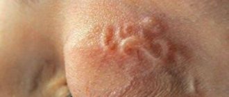
With this type of disease, both the superficial layer of the skin (epidermis) and the connective tissue layers are affected. Often the disease occurs like a boil, differing only in migration from one area of the body to another.
Symptoms of cutaneous (subcutaneous) myiasis:
- the appearance of a moving subcutaneous swelling;
- itching and burning in the area of swelling;
- the appearance of an ulcer at the site of swelling;
- allergic reaction in the area of swelling.
With timely treatment, the prognosis is favorable, but in its absence, severe and deep ulcers, as well as severe allergic reactions, may occur.
Treatment: surgical removal of larvae from the skin followed by treatment of the wound with antiseptics.
Ocular myiasis (ophthalmomyasis)
Symptoms of ocular myiasis (ophthalmomyasis):
- pain in the eyeball;
- visual impairment (decreased vision in the affected eye, appearance of floaters, double vision);
- lacrimation, redness of the affected eye.
An extremely dangerous form of the disease, often leading to loss of vision or even death.
A relatively rare form of the disease, usually found in people without a fixed place of residence or in animals.
Symptoms of nasal myiasis:
- pain (including bursting) in the nasal passage;
- copious nasal mucus;
- sneezing;
- redness of the skin of the nose.
Due to its proximity to the eyes and brain, this form of the disease is considered dangerous, so treatment should be started immediately.
Infection with ear myiasis in the vast majority of cases occurs through the passive participation of a person, usually during sleep. Drug therapy and rinsing for this form of the disease are pointless; the only treatment method is surgery.
Symptoms of ear myiasis (otomiasis):
- bursting pain in the ear canal;
- congestion in the affected ear, hearing loss;
- inflammation of the affected ear (ordinary touching it causes pain).
This form of the disease is dangerous because the larvae can bore passages from the affected ear to the brain, which can result in meningitis, thrombosis of the venous sinuses and, accordingly, the death of the patient.
Myiases mean all diseases caused by the larvae of many species of flies. Simply put, myiases are an umbrella term that includes many diseases. But what specific diseases do fly larvae cause?
Fly larvae lead to the development of the following diseases in humans and animals:
- Wolfarthiosis. The causative agent is the larva of the Wohlfarth fly. The disease affects humans and animals, leading to unbearable pain, tissue necrosis and often death.
- Gastrophilosis. The causative agent is the larva of the gadfly Gastrophilus equi. The disease affects mammals (including humans), leading to extensive damage to various organs.
- Hypodermatosis. The causative agent is the larvae of the gadfly Hypoderma bovis and Hypoderma lineatum. The disease affects humans and cattle, leading to the formation of ulcerative diseases of the gastrointestinal mucosa.
- Dermatobiasis. The causative agent is the larva of the gadfly Dermatobia hominis. The disease affects humans and animals, leading to the development of local inflammation and abscesses.
- Cordylobiosis. The causative agent is the larva of the fly Cordylobia anthropophaga. The disease affects animals and humans, leading to the development of abscess nodes, ulcers and boils (especially in children).
Treatment of myiases
In the vast majority of cases, myiases are treated with medications aimed at destroying the larvae, reducing inflammation and eliminating secondary infections. For these purposes, such drugs as “Mebendazole”, “Penicillin”, “Difezil” and so on are used.
Surgical intervention is also extremely effective, in which fly larvae and necrotic (literally dead) body tissues are removed from the affected organs. In addition, during surgical intervention, purulent masses are drained from the site of larval penetration.
Surgical treatment is used both as an analogue to drug treatment, if the use of the second is pointless or difficult, and as a primary treatment to achieve rapid subsidence of inflammatory and allergic phenomena.
For reference!
When the larva moves to the next stage, it no longer poses any danger to humans.
Infection with cutaneous myiases can occur in several ways:
Oral (dental) myiasis is diagnosed less frequently than cutaneous myiasis, but it can also cause problems for a person. The larva enters the oral cavity along with food. The disease is accompanied by symptoms such as:
- constant itching in the mouth;
- swelling of the gums;
- nasal congestion;
- long continuous cough;
- general malaise;
- increased body temperature;
- fever;
- bleeding gums.
The disease is caused by the oral route and is caused by the larvae of the Wohlfarth fly. The risk group includes patients who do not follow the rules of personal hygiene, for example, do not brush their teeth at least once a day. The disease can also affect people who have purulent wounds in the mouth, alcoholics, and the elderly.
Intestinal myiasis is diagnosed when the larva first enters the oral cavity and then moves to the gastrointestinal tract. When the intestines become infected, the patient experiences severe abdominal pain.
Intracerebral infection is extremely rare. The symptoms of this disease are slightly different from the usual disease. The patient is worried about convulsive conditions, high body temperature, back pain, and the appearance of boils.
Important!
To cure intracerebral myiasis, surgery is necessary. The surgeon will mechanically remove all larvae.
Types of myiasis[edit | edit code]
Depending on the affected organ or tissue, there are cutaneous, genitourinary (urinary), intestinal myiases, nasal (nasal) myiases, otomiasis (ear myiases), ophthalmomyasis (eye myiases).
Ophthalmomyasis
- severe eye damage caused by larvae of gadflies and Wohlfarth flies. Larvae, lingering in the thickness of the conjunctiva, contribute to the development of chronic conjunctivitis; they can penetrate through the limbus into the anterior chamber, into the vitreous body, leading to severe iridocyclitis. The process may result in the loss of an eye.
Varieties of myiases
Ocular, or ophthalmic myiases - the larvae penetrate directly into the eyes, resulting in the formation and accumulation of pus and, as a consequence, conjunctivitis. With an advanced form of the disease, retinal detachment may begin, ulcers appear on the cornea, and accordingly, vision deteriorates, and sometimes the eye is simply eaten.
If the larvae penetrate the brain, encephalitis begins. Larvae in the nasal cavity provoke rhinitis, in the auricle - destruction of the eardrum and parasitism on the membranes of the brain.
Cavity myiases - penetration of larvae into body cavities, resulting in purulent formations, multiple ulcers and perforations, destruction of organ walls. In particular, intestinal myiases can manifest as colitis, enteritis, diarrhea, vomiting and other symptoms. When parasitizing the mucous membranes of the genitourinary organs, problems with urination, cystitis, vulvitis, etc. usually occur.
Diseases[edit | edit code]
Wolfarthiosis
- an invasive disease of animals and humans caused by the larva of the Wohlfarth fly when it develops in wounds, macerated skin or on the mucous membranes of natural orifices.
Wohlfahrtia magnifica
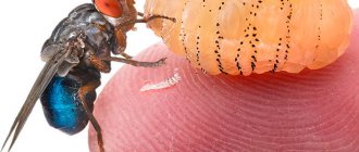
is common in temperate and hot climates. The body of the fly is light gray in color and has a length of 9-13 mm. Adult flies live in fields and feed on plant nectar. Female flies hatch from 120 to 150 larvae in open cavities (nose, eyes, ears), on wounds and ulcers on the body of animals, sometimes on humans (while sleeping in the open air). Larvae in humans live in the ears, nose, frontal sinuses, and eyes. Having quickly penetrated tissues, the larvae destroy them down to the bones mechanically and with the help of secreted enzymes. Parasitization of larvae is accompanied by severe pain, causes tissue necrosis and gangrenous processes. After 5-7 days, the larvae fall into the soil and pupate. After 11-23 days, an adult insect develops from the pupa in the soil. By making passages in the tissues, the larvae not only cause painful sensations - the damaged areas swell and fester, the tissues partially die, and bleeding begins from the nose. Myiasis is very painful and often ends in death. After removing the larvae, all these phenomena disappear. At one time, a Wohlfarth fly can lay 120-160 pieces. larvae, larvae up to 1 mm long. Females live 8-29 days.
Obligate myiases
Malignant myiases
are caused mainly by the larvae of the Wohlfart fly (Wohlfahrtia magnibica), common in Southern Europe, the Arab Republic of Egypt, Mongolia, China, the Caucasus, Central Asia, Kazakhstan, the central and southern zones of Russia.
The fly lays 120-160 very mobile larvae, about 1 mm long, on the skin of humans and animals. They quickly penetrate through the skin deep into the tissues, down to the bones, and move into the eyes, nose, ears and even the brain, exerting a mechanical, allergoid and enzymatic effect on the tissue. In a person infested with Wohlfarth fly larvae, necrosis and suppuration occur, and extensive destruction of tissues and organs occurs. Cases of complete destruction of the eyeball by larvae, destruction of the scalp, and the occurrence of osteomyelitis, encephalitis, and severe damage to the female genital organs under their influence are described. Fatalities are known. The disease progresses with extraordinary speed.
Wohlfarth fly larvae develop very quickly: after 3-5 days they become mature, leave the host and pupate in the soil.
Treatment of malignant myiasis consists of washing wounds with chloroform water or irrigating with a solution of chloroform in vegetable oil, removing euthanized larvae with tweezers, removing necrotic tissue, opening abscesses, using antibacterial drugs, and administering antitetanus serum.
The Wohlfarth fly lays larvae primarily on ungulates. In order to prevent and combat malignant myiasis, periodic examinations of farm animals (sheep, etc.) and removal of fly larvae are of great importance.
Benign myiases
are caused by larvae that develop singly and slowly. There are African and South American myiases.
Infection[edit | edit code]
Female flies lay eggs in the eyes, ears, nose, wounds of people, or inject them subcutaneously. Less common is visceral damage caused by accidental ingestion of larvae[3]. For example: representatives of the genus Gasterophilus
,
Hypoderma
,
Dermatobia
and
Cordylobia
affect the skin;
Fannia
affects the gastrointestinal tract and urinary system;
Phonnia
and
Wohlfahrtia
can infect open wounds and ulcers;
Oestrus
affects the eyes;
as well as Cochliomyia
penetrate the nasal passages and carry out their invasion.
South American myiasis
South American myiasis occurs in Mexico, Argentina and other countries in Central and South America. The causative agent is the larva of the fly Dermatobia hominis. The female fly lays eggs on the body of mosquitoes, jet flies and some ticks. After 6 days, larvae form in the eggs, but they leave the egg shells only when the insects and mites on which they are located land on humans or animals (cattle, pigs, etc.). The larvae quickly penetrate the skin, where they slowly grow and develop. After 5-10 weeks they reach 25 mm in length, fall out onto the soil and pupate.
An infiltrate with a hole appears around the embedded larvae, from which serous-purulent fluid flows. Lesions are located mainly on the limbs, back, abdomen, and armpits. A case of death of a 1.5-year-old child with multiple infestations by the larvae of this fly is described.
Treatment is carried out by removing the larvae with tweezers.
Prevention is carried out through routine examination of domestic ungulates and treatment of patients, control of blood-sucking insects and ticks using repellents.
Human defeat[edit | edit code]
The main causative agent is the tumbu fly ( Cordylobia anthropophaga
).
Wohlfahrtia magnifica
fly and various gadflies, most often the sheep gadfly (
Oestrus ovis
)[3].
There are three main groups of flies that can cause myiasis in humans:
- Calliphorid family ( Calliphoridae
) - Superfamily Oestroidea
- mainly a family of gadflies (
Gasterophilidae
) - Gray blowfly family ( Sarcophagidae
)
Other groups that, although less frequently, can also cause myiases:
- Centipede family ( Anisopodidae
) - Cheese fly family ( Piophilidae
) - Lionfly family ( Stratiomyidae
) - Hoverfly family ( Syrphidae
)
Types of flies whose larvae require a host:
- Dermatobia hominis
- Cordylobia anthropophaga
- Oestrus ovis
- Hypoderma
sp. - Gasterophilus
sp. - Cochliomyia hominivorax
- Chrysomya bezziana
- Auchmeromyia senegalensis
- Cuterebra
sp.
Links[edit | edit code]
- Exotic Myiasis (English). Department of Medical Entomology, University of Sydney (medent.usyd.edu.au). Access date: July 17, 2013. Archived August 29, 2013.
- Blowfly strike in sheep (English). cahl.ie; archive.org. — The inaccessible link has been replaced with an archived one. Access date: July 17, 2013.
- Ramli R., Rahman RA
Human oral myiasis, two cases and general remarks (English).
Malaysian Journal of Medical Sciences, Vol.
9, No. 2, July 2002, pp. 47–50 . bioline.org.br (2002). Access date: July 17, 2013. Archived August 29, 2013. - Jiang C.
A collective analysis on 54 cases of human myiasis in China from 1995-2001 (English) (PDF).
Chin Med J (Engl).
2002 Oct;115(10):1445-1447 . Chinese Medical Journal. Access date: July 17, 2013. Archived August 29, 2013. - Identification key to myiasis causing fly larvae. Nat Hist Museum (nhm.ac.uk). Access date: July 17, 2013. Archived August 29, 2013.

