Most people consider helminthiases to be primarily an intestinal disease, but there are types of worms that parasitize other human organs, even the brain. Worms in the human brain cause great harm to the entire body, and in particularly advanced cases can even lead to death.
Let's look at two main diseases caused by parasitism of worms in the brain, which are more common in our country - neurocysticercosis and echinococcosis; Let's figure out how worms get into the head, how these diseases are diagnosed, how they are treated and what can be done to prevent them.
How parasites get into the human brain
Cysticercosis
Cysticerci are pig tapeworm eggs, which, after entering the gastrointestinal tract, can invade the human nervous system. The adult lives in the intestines, but the parasite constantly multiplies, releasing a huge number of eggs.
They enter the stomach and intestines; in the stomach, under the influence of the digestive process, the protective shell of the eggs dissolves, and as a result, small larvae begin to circulate throughout the body along with the bloodstream.
In rare cases, the parasite larvae establish themselves in certain other organs, such as skeletal or eye muscles, and they can also infect the brain.
As practice has shown, in approximately 6 out of 10 cases, the larvae settle in the head, and not other organs; here they can exist for up to three decades, causing a dangerous disease - neurocysticercosis.
The cause of the disease is T. solium (pork tapeworm). The source of the causative agent of the invasion is a sick person who excretes mature eggs and tapeworm segments with feces. Infection occurs through contaminated hands, food, water, as well as through the consumption of pork that has undergone insufficient heat treatment, for example, poorly fried kebabs.
Pork tapeworm
Cysticercosis develops as a result of pork tapeworm eggs entering the stomach with contaminated products, through dirty hands, when mature segments are thrown from the intestines into the stomach during vomiting in persons infected with the sexually mature form of pork tapeworm.
If a person swallows an egg, the stomach and duodenum release larvae, which enter the bloodstream through the intestinal wall and spread throughout the body. In the place where the larva attaches (for example, in the muscles or in the human brain), it turns into a cysticercus, which matures within 2-3 months and lives for several years.
Cysticercus in the brain
A cysticercus is a pea-sized to walnut-sized bubble filled with clear liquid (from 3 to 15 mm in diameter).
On the inner surface of the bladder is the head of the finna - the scolex with hooks and suction cups. In most cases, there are hundreds and thousands of parasites in the brain, but single cysticerci are also found.
They are localized in the soft meninges at the base of the brain, in the superficial parts of the cortex, in the cavity of the ventricles, where they can float freely.
When the parasite dies, it becomes calcified, however, remaining in the brain, it supports a chronic inflammatory process. The lifespan of the parasite is from 5 to 30 years.
Echinococcosis
Echinococci are tapeworms that parasitize the intestines of dogs, wolves, jackals, and less commonly cats. Humans are their intermediate host at the larval stage. Echinococcus larvae are dangerous human parasites that cause echinococcosis. Due to its slow development and life cycle, the parasite manages to cause irreparable damage to the internal organs in which it is localized.
Infection occurs orally. In the intestine, a larva emerges from the egg with six movable hooks at the posterior end of the body (oncosphere).
With the help of hooks, it penetrates through the intestinal wall into the circulatory system and is transported with the blood to the liver or other organs, including the brain. Here the oncosphere develops into the vesicular stage (finna), which is also called echinococcus.
Echinococcus
The oncosphere, like the cysticercus, forms a bubble; secondary and even tertiary bubbles appear on its walls, on which many scolex are formed. Echinococcus blisters grow very slowly and can reach the size of a child's head.
You can become infected with echinococci through contact with sheep and dogs, as well as wild animals; failure to observe personal hygiene rules, eating unwashed food and dirty water.
Oncosphere
What is neurocysticercosis?
A disease in which tumors with worms inside form in the human brain is called neurocysticercosis . It is caused by the tapeworm Taenia solium, which is also known as the pork tapeworm. The length of an adult individual can reach 3 meters, and on the head there are suction cups, with the help of which it is attached to the intestines of animals. The majority of carriers of these parasites are pigs.
Worms do not enter the brain immediately - for this to happen, special conditions are needed. First, the eggs of the pork tapeworm enter the animals' bodies through the ground, accumulate in the meat, and the larvae hatch from them. When a person eats contaminated meat, the larvae enter the intestines and eventually grow, causing a disease called taeniasis - symptoms include weakness, nausea and cramping abdominal pain. This disease is treated with the medicine fenasal or male fern extract, which kill parasites and remove them from the body.
Symptoms of worms in the human brain
Symptoms of neurocysticercosis can be very different and depend on where exactly the cysts are localized, as well as on their number and stage of development. In total, it is customary to distinguish 6 different forms, each with its own symptoms. In general, the clinical signs of the disease are characterized by:
- Mental changes;
- Paresis and paralysis;
- Visual impairment and breathing disorders;
- Headaches, sometimes with symptoms of meningitis, nausea and vomiting;
- Convulsions, epileptic seizures, etc.
In addition to the toxic and sensitizing effects of enzymes and metabolites of larvae during migration, an important factor in pathogenesis is the mechanical pressure of the growing parasite on surrounding tissues, which causes severe headaches.
Echinococcosis in its symptoms resembles a tumor with severe hypertension syndrome. The following symptoms are observed:
- Increased intracranial pressure.
- Dizziness and constant headaches;
- Attacks of vomiting;
- Epileptic seizures;
- Congestive discs of the nerves of the visual organs;
- Blood eosinophilia;
Are maggots useful?
The positive effects of worms on trophic and purulent infected wounds have been clinically proven.
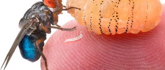
Blowfly larvae eat decaying tissue. Their digestive system has external development. Once in the wound, they release proteolytic enzymes that help dissolve dead cells. Natural cleansing of necrotizing lesions occurs. The use of maggots can be compared to surgical practice, where pancreatic enzymes are used. Trypsin is placed into the pathological focus, which cleanses the wounds of purulent contents. The main advantage of larval therapy is its rapid and long-term effect.
Scientists are developing an antibiotic that can help with non-drug cases, gangrene. The substance isolated from the processed secretion of green fly larvae was named Seraticin. It has shown effectiveness in the treatment of diseases caused by E. coli, staphylococci, and clostridia.
Another positive effect of maggots on a wound is the suppression of local immunity. The increased protective reaction of the skin causes typical symptoms of inflammation. The patient suffers from swelling and pain. Thanks to mucus, the larvae relieve swelling, reduce hyperemia, and fight pain.
The therapeutic effect of worms is manifested in many difficult situations: bedsores, non-healing trophic ulcers associated with metabolic disorders in diabetes, poor circulation of the lower extremities, atherosclerosis, improper functioning of the nervous system, various types of polyneuropathy. The main use of blowfly maggots on a wound is the fight against dry and wet gangrene. Antibiotics are often powerless against suppurative processes on the extremities. Gangrene can only be treated surgically. Larval therapy will help preserve the limb if used in a timely manner.
Diagnostics
Neurocysticercosis is one of the most dangerous diseases, including because it may not manifest itself in any way for a long period. Diagnosing neurocysticercosis is quite difficult, since each of the symptoms individually is not specific for neurocysticercosis. Neurocysticercosis should be distinguished from diseases that have similar symptoms: tumor, epilepsy, neurosyphilis, meningoencephalitis and others.
When making a diagnosis, the totality of symptoms is important: a multiplicity of symptoms as a result of multiple lesions of brain formations, the manifestation of symptoms of irritation, symptoms of increased intracranial pressure, alternation of a severe condition and light intervals with the absence of any signs of cysticercosis. The diagnosis is confirmed by instrumental and laboratory research methods
The main diagnostic method is a blood test for eosinophils. It is the increased level of these cells in the blood that allows one to suspect the presence of parasites. Then an analysis of the cerebrospinal fluid is performed, in which antibodies can be found, and an X-ray of the head is done - here you can see calcifications. A more detailed picture of the disease will be shown by CT or MRI, as well as radiographic indicators.
A study of spinal fluid is informative. For this, a spinal tap is performed. An increase in the content of lymphocytes and eosinophils and protein content is detected in the cerebrospinal fluid. In rare cases, it is possible to detect parts of the scolex and cysticercus capsules. The puncture is performed only by a doctor, usually an anesthesiologist, in a hospital setting under aseptic conditions.
RSC of the blood or cerebrospinal fluid is performed using a specific cysticercosis antigen. The Lange reaction is paralytic.
CT and MRI scans of the brain reveal varying numbers of small formations that have dense contours.
With echinococcosis, timely diagnosis is crucial. Slowness in such situations can cost a person his life, since echinococcosis cysts can quickly develop into tumors. The growth of cysts occurs quite quickly, causing pathological and mechanical damage.
Diagnostic methods for echinococcosis:
- Ultrasound of all internal organs in the peritoneum and retroperitoneal space;
- Echography;
- Blood and urine tests, general and biochemical blood tests;
- Blood test for antibodies;
- X-ray of all organs in the chest area;
- Magnetic resonance and computed tomography;
- ERCP;
- Spirography.
In most cases, echinococcosis is diagnosed completely by accident during an examination for a completely different reason, and immediate initiation of treatment gives hope for a favorable outcome.
Corpse fauna
The housing problem can spoil not only people, but also worms. As soon as a fresh living space looms on the horizon, parasites will immediately populate the square meters without asking, so that they don’t sit idle unnecessarily. There is some truth in this joke, but it lies deeper.
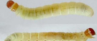
The fact is that dead biological tissue attracts lower insects, which in this case play the role of orderlies. Where do maggots come from in humans? It must be said that such an unpleasant fate is inevitable for dead bodies, that is, dead people. Here, in principle, everything is clear - this process is natural and does not require explanation.
But sometimes out-of-the-ordinary things happen - maggots in a person... alive! It turns out that such cases, although rare, do occur. Moreover, it is impossible to insure against them.
One of the reasons for the appearance of parasites may be a bleeding wound or advanced gangrene. Particularly dangerous, as you might guess, is the summer period, when there are especially many flies. It is enough to go to bed, leaving access to the wound, and by the evening of the next day, strange white dots may appear on the damaged bleeding area... Only careful treatment, applying a bandage and bandaging can save it.
Maggots can also settle in humans thanks to food. It turns out that the peak breeding activity of the gray fly occurs either at the end of summer or at the beginning of autumn. At this time, the insect lays its eggs on the food with special zeal. According to some entomologists, after a person eats a contaminated product, the larvae get inside and some of them survive.
Parasites continue their development inside. They come out with feces, but during this time, in addition to unpleasant sensations, the person receives ascaris eggs as a “gift”, which are carried by flies on their paws. In order to convince you to thoroughly wash or heat-treat food, it is probably enough to say that one such parasite reaches forty centimeters in length and lays up to two hundred eggs in just one day!
Treatment
Neurocysticercosis can last for years in an erased form, without revealing itself in any way. After clarifying the diagnosis, only a parasitologist or infectious disease specialist can prescribe a course of deworming.
Treatment of neurocysticercosis is carried out with medication and surgery. It must be individual and selected based on the location and number of cysticerci, as well as the characteristics of the host’s immune response.
Drug therapy consists of the use of antiparasitic drugs, which include Nemozol, Azinox, Cestox, Paraziquantel, Sanoxal, Albendazole. Their action is aimed at destroying parasites. However, their breakdown products have a toxic effect on the surrounding brain tissue.
Symptoms may worsen after using these medications. Therefore, anti-inflammatory drugs and, if necessary, hormonal drugs are added to the treatment. Anti-edematous therapy is carried out. If necessary, antiemetics and painkillers are prescribed.
If there are single cysts, as well as if cysticerci are located in the IV ventricle, or in relatively easily accessible areas of the cerebral cortex, then surgical intervention is performed to remove them. This intervention can sometimes lead to recovery. With numerous cysts, such removal is impossible, and the prognosis for life is very unfavorable.
But more often it is not possible to remove all cysticerci. Some may go completely unnoticed. Therefore, surgical treatment is supplemented with medicinal anthelmintic drugs.
For echinococcosis, the main treatment method is surgical removal of the hydatid cyst followed by antiparasitic therapy.
Currently, the standard of neurosurgery for echinococcosis of the brain is the method of microsurgical preparation and removal of cysts with an intact capsule, since when a cyst ruptures, the surrounding tissues are contaminated with protoscolex of the parasite, and anaphylactic shock often develops. When cysts are localized in functionally significant areas of the brain, the use of intraoperative neurophysiological monitoring is mandatory.
When carrying out conservative therapy, the most effective and generally accepted drug is albendazole (andazole, escazole, nemozol, zentel), which irreversibly disrupts the utilization of glucose by parasites, which leads to their death.
Chemotherapy was previously used to treat inoperable patients, but its indications have now been expanded.
Prevention
The simplest measures to prevent both neurocysticercosis and echinococcosis are:
- Compliance with generally accepted rules of hygiene, especially when contacting domestic animals and processing carcasses and skins of killed animals;
- Thorough heat treatment of food products, especially meat, as they may contain parasite larvae;
- Thorough washing of fruits, berries, vegetables consumed raw;
- Refusal to use water from springs, streams, wells without prior boiling.

