Etiology
The causative agent, Rickettsia sibirica, was discovered in 1938 by O.S. Korshunova. Like other pathogens of the group of spotted fevers, it parasitizes both in the cytoplasm and in the nucleus of the affected cells. Antigenically it differs from other rickettsiae in this group. Contains a toxic substance. Characterized by properties common to all rickettsiae. Able to persist for a long time in the external environment at low temperatures (up to 3 years). Keeps well when dried. The virulence of individual strains varies significantly.
The causative agent of tick-borne typhus
Tick-borne typhus is caused by pathogenic microorganisms from the rickettsia group. Their specific species is Rickettsia sibirica. It has common properties characteristic of all representatives of rickettsia. The only difference is moderate virulent abilities. Therefore, its entry into the body does not cause severe manifestations.
According to the morphological structure, Rickettsia sibirica is a gram-negative rod with an aerobic type of metabolism. The only natural reservoir for it is the rodent organism. Ixodid ticks act as a carrier of infection, which ensures its constant circulation in a certain area. This type of rickettsia is very resistant to high and low temperatures in the external environment. Different strains may have different virulent and pathogenic properties, determining the clinical course of the disease.
In most cases of Rickettsia sibirica disease, it is timely verified by the body's immune cells. Its destruction does not release dangerous endotoxins. This allows the body to cope with the pathogen on its own, even in the absence of treatment. As a result, persistent immunity occurs in the form of antibodies to the antigenic components of this type of rickettsia, which remains for life.
Epidemiology
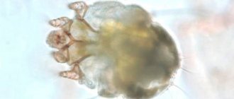
The disease is classified as a zoonotic disease with natural focality. Natural foci have been identified in the Primorsky, Khabarovsk and Krasnoyarsk territories, in a number of regions of Siberia (Novosibirsk, Chita, Irkutsk, etc.), as well as in Kazakhstan, Turkmenistan, Armenia, Mongolia. The reservoir of rickettsia in nature is about 30 species of various rodents (mice, hamsters, chipmunks, gophers, etc.). Transmission of infection from rodent to rodent is carried out by ixodid ticks (Dermacentor nuttalli, D. silvarum, etc.). The infestation of ticks in foci reaches 20% or more. The incidence in the tick habitat ranges from 71.3 to 317 per 100,000 population per year. The immune layer of the population in natural foci ranges from 30 to 70%. Rickettsia remain in ticks for a long time (up to 5 years); transovarial transmission of rickettsia occurs. Not only adult ticks, but also nymphs participate in the transmission of infection to humans. Rickettsia is transmitted from ticks to rodents through blood sucking.
A person becomes infected during his stay in the natural habitats of ticks (shrubs, meadows, etc.), when he is attacked by infected ticks. The greatest activity of ticks is observed in spring and summer (May-June), which determines the seasonality of the incidence. The incidence is sporadic and occurs mainly among adults. Not only rural residents get sick, but also those traveling outside the city (garden plots, recreation, fishing, etc.). In recent years, about 1,500 tick-borne rickettsiosis diseases are registered annually in Russia.
Lenin's bagmen
Dramatic events unfolded in Russia. The territory controlled by the Soviet government was gripped by an unprecedented epidemic of typhus. Previously, this disease was rampant only in a war zone or in a single city. But now it has spread everywhere. The bag men carried it away. Since Lenin banned the sale of food products in stores and the transportation of flour by wagons, supplies came only through private speculators. They reached the Ukrainian border or the front line, behind which food was found, and there they bought bread, flour and cereals. Bag makers moved illegally in freight cars that no one disinfected. The trouble is that the incubation period of typhus is at least 5 days, and during this time the patient can travel very far.
People's Commissar of Health of the RSFSR Nikolai Semashko explained to the population the role of lice and called on them to fight it. But that was all there was to it, since the Russian chemical industry was gone and there was no import of disinfectants. Soviet citizens mockingly nicknamed body lice “semashkas.” The expression “to catch a poisonous haemorrhage” meant to be bitten by typhoid lice. There was nothing to disinfect medical institutions with. People with typhus were placed in the corridors in their outer clothing - that is, in Tunisia the situation was better. In Moscow, almost all the doctors became infected, half died, especially the elderly and those with weak hearts. The population was left alone with typhus, and during the civil war, 30 million people were ill.
Pathogenesis
The portal of infection is the skin at the site of the tick bite (rarely, infection occurs when rickettsia is rubbed into the skin or conjunctiva). At the site of introduction, a primary affect is formed, then rickettsiae move along the lymphatic pathways, causing the development of lymphangitis and regional lymphadenitis. Lymphogenously, rickettsiae penetrate into the blood and then into the vascular endothelium, causing changes of the same nature as in epidemic typhus, although they are much less pronounced. In particular, there is no necrosis of the vascular wall, thrombosis and thrombohemorrhagic syndrome rarely occur.
Endoperivasculitis and specific granulomas are most pronounced in the skin and to a much lesser extent in the brain. Allergic restructuring is more pronounced than in epidemic typhus. The transferred disease leaves a strong immunity, recurrent diseases are not observed.
Mochutkovsky's experiment
The experiment made such a strong impression on the infectious disease specialist that in 1876 Mochutkovsky performed the same one on himself, but with typhus. He almost died, developed myocarditis, migraines and memory loss. And yet the result was achieved: the typhus clinic is obvious, but there are no spirochetes in the blood. Therefore, real typhus - the menace of any army - is caused by something else, and this something can be carried by insects. Which ones? In a historical letter to the editor of the Chronicle of Medicine dated February 2 (14, new style), Minkh suggested that researchers “collect a small number of known insects (bugs, fleas...), which are easy to find...”
The hypothesis caused distrust: since both work in a hospital, they could have become infected from patients. Then the biologist Ilya Mechnikov, who did not communicate with patients by nature of his work, inoculated himself with relapsing fever and suffered from it (biographers see in this feat a disguised suicide attempt), proving Minha right: blood is contagious.
Symptoms and course
The incubation period ranges from 3 to 7 days, rarely up to 10 days. There are no prodromal phenomena (with the exception of the primary affect, which develops soon after a tick bite). As a rule, the disease begins acutely, with chills, body temperature rises, general weakness, severe headache, pain in muscles and joints appear, sleep and appetite are disturbed. Body temperature in the first 2 days of illness reaches a maximum (39-40°C) and then persists as a fever of a constant type (rarely remitting). The duration of fever (without antibiotic treatment) most often ranges from 7 to 12 days, although in some patients it lasts up to 2-3 weeks. When examining the patient, mild hyperemia and puffiness of the face are noted. Some patients experience hyperemia of the mucous membrane of the soft palate, uvula, and tonsils. The most typical manifestations are primary affect and exanthema. When bitten by uninfected ticks, the primary affect never develops; its presence indicates the onset of the infectious process.
The primary affect is an area of infiltrated moderately compacted skin, in the center of which necrosis or a small ulcer covered with a dark brown crust is visible. The primary affect rises above the skin level, the zone of hyperemia around the necrotic area or ulcer reaches up to 2-3 cm in diameter, but there are changes of only 2-3 mm in diameter, and they are quite difficult to detect.
Not all patients note the very fact of a tick bite. Healing of the primary affect occurs after 10-20 days. In its place there may be pigmentation or peeling of the skin. A characteristic manifestation of the disease is exanthema, which is observed in almost all patients. It usually appears on days 3-5, rarely on days 2 or 6 of illness. First it appears on the limbs, then on the torso, face, neck, and buttocks. The rash is rarely observed on the feet and palms. The rash is abundant, polymorphic, consists of roseola, papules and spots (up to 10 mm in diameter).
Hemorrhagic transformation of rash elements and the appearance of petechiae are rarely observed. Sometimes there is a “sprinkling” of new elements. The rash gradually disappears by 12-14 days from the onset of the disease. There may be peeling of the skin at the site of the spots.
In the presence of primary affect, it is usually possible to detect regional lymphadenitis. Lymph nodes are enlarged to 2-2.5 cm in diameter, painful on palpation, not fused to the skin and surrounding tissues, suppuration of the lymph nodes is not observed.
From the cardiovascular system, bradycardia is noted, a decrease in blood pressure, arrhythmias and changes in the heart muscle according to ECG data are rare. Changes in the central nervous system are observed in many patients, but do not reach the same extent as occurs with epidemic typhus.
Patients are bothered by severe headaches, insomnia, patients are inhibited, agitation is observed rarely and only in the initial period of the disease. Mildly expressed meningeal symptoms are very rarely detected (in 3-5% of patients); when examining cerebrospinal fluid, cytosis usually does not exceed 30-50 cells in 1 μl. There are no pronounced changes in the respiratory system. Enlargement of the liver is observed in half of the patients, the spleen increases less frequently (in 25% of patients), the increase is moderate. The course of the disease is benign. After the temperature drops to normal, the patients' condition quickly improves and recovery occurs quickly. Complications, as a rule, are not observed. Even before the use of antibiotics, the mortality rate did not exceed 0.5%.
Medical reference books
anthrax
ICD-10: A22
anthrax
– an acute infectious disease caused by the pathogen Bac. anthracis from the group of zoonoses, characterized by fever, damage to the lymphatic system, and intoxication.
Historical data
Anthrax has been known to mankind for a long time. Ancient manuscripts have been preserved in which the disease is described under the name “sacred fire”, “Persian fire”. Significant epidemic outbreaks of anthrax were noted in Europe and Siberia in the 17th century. S.S. Andreevsky, in the experience of self-infection, established the identity of anthrax in humans and animals. The domestic veterinarian F. Browell is credited with the discovery of the causative agent of anthrax in the blood of a deceased person. The creation of the doctrine of anthrax is closely connected with the names of R. Koch, L. Pasteur, I.I. Mechnikova, L.S. Tsenkovsky, N.F. Gamaleya. In 1876, R. Koch isolated the causative agent of anthrax in pure culture, grew it on an artificial nutrient medium, detected sporulation and reproduced the infection in an experiment on mice. L. Pasteur proved that the pathogen in the form of spores persists in the soil for many years and recommended burning the corpses of sick animals. He was the first to propose a live vaccine for the specific prevention of anthrax.
Etiology
The causative agent of anthrax is a gram-positive bacterium (Bac. anthracis) of fairly large size (5-8 microns in length and 1.5-2 microns in width), with characteristic “chopped off” ends. Aerobes easily form spores in the external environment, which are highly resistant to physical and chemical influences. Anthrax spores die at a temperature of 120 °C only after 2 hours.
Epidemiology
Source
Anthrax for humans are animals that are sick or have died from it. Transmission of the pathogen from an animal to a person is most often carried out by contact through direct contact with a sick animal, with contaminated raw materials of animal origin, with finished products, or soil. Infection can also be carried out through nutritional, vector-borne and airborne routes. The disease is usually registered in the summer-autumn months, which is associated with a seasonal increase in the incidence of farm animals. The disease affects mainly men of active working age (20-50 years), who more often than women have to participate in caring for sick domestic animals, slaughtering them, cutting up carcasses, etc.
Pathogenesis
A reflection of previous ideas about the pathogenesis of human anthrax is the division of clinical forms of anthrax into cutaneous, intestinal, pulmonary and septic. Currently, there are only two clinical variants of the disease: localized and generalized. Localized anthrax includes the cutaneous form. In the cutaneous form, there must be skin changes at the entrance gate. Generalized anthrax is a consequence of the pathogen entering the blood from the lymphatic system. The entry points for infection are the skin, the mucous membrane of the respiratory tract and, rarely, the mucous membrane of the gastrointestinal tract. The pathogenesis of anthrax appears to be a two-stage process. Regardless of the entrance gate of the infection, the first stage represents a localized lesion of regional lymph nodes, the second stage is a generalization of the process.
The pathogenesis is based on the action of the pathogen exotoxin, which consists of at least three components or factors: the first (I) - edema (inflammatory) factor, the second (II) - protective (protective) antigen (RA) and the third (III) – lethal factor. Adding factor I to factor II increases the immunogenic properties, and adding factor III reduces them. A mixture of factors I and II causes an increase in the inflammatory response and edema due to an increase in capillary permeability. A mixture of factors II and III enhances the effect of the lethal factor and leads to the death of guinea pigs, rats and mice. A mixture of three anthrax toxin factors (I, II, III) has an inflammatory and lethal effect.
A person most often becomes infected through damaged skin and aerogenic (inhalation) routes. Other methods of infection rarely lead to illness. In some cases, the disease occurs in the form of localized anthrax and the formation of a skin carbuncle, in others - in the form of a general infection, ending in generalization of the process.
When infected through the respiratory tract, and in some cases through damaged skin and mucous membrane of the gastrointestinal tract, the process can immediately develop according to the type of generalization and be very acute.
The entry point for cutaneous anthrax can be any part of the skin and mucous membranes. It can be considered that the cutaneous form is caused, first of all, by a small dose of infecting material entering the skin and its superficial penetration. An anthrax carbuncle develops at the site where the pathogen enters the skin.
- a peculiar skin lesion inherent in humans and representing a focus of serous-hemorrhagic inflammation with necrosis, swelling of surrounding tissues and regional lymphadenitis. In the edema zone, the patency of the lymphatic vessels is completely preserved both in the early and late stages of the development of edema. This makes it possible for the anthrax pathogen to be introduced from the site of penetration by mobile macrophages along the lymphatic tract into the lymph nodes, in which serous, serous-hemorrhagic or necrotic-hemorrhagic inflammation develops. The causative agent of anthrax lingers in the lymph nodes for some time, part of it dies, and the part that remains viable enters the general bloodstream. The aerogenic route of infection is possible only through spores. anthracis when they are transferred together with dust into the air (in an aerosol state). Spores can easily be converted into an aerosol state during various technological operations when processing dry infected animal raw materials: skin, wool, hair, bristles, etc. The higher the concentration of aerosol particles loaded with spores, the greater the risk of infection. Infected aerosol particles usually settle on the mucous membrane of the trachea and bronchi, without reaching the alveoli. Anthrax bacillus spores do not germinate on the epithelium of the trachea and bronchi. A significant part of the pathogen is removed from the lungs with secretory secretions. Some of the pathogen is captured by mobile macrophages and carried through the lymphatic ducts to regional (mediastinal, tracheal, bronchial) lymph nodes, in which they germinate and multiply, which is accompanied by an inflammatory reaction of the lymph nodes and their destruction. Hence, the branched lymphatic and circulatory system allows the pathogen to quickly and in massive doses break into the bloodstream. This is facilitated by the partially closed lymphatic system of the lungs, where the pathogen receives unlimited opportunities to reproduce and enter the blood. In the initial period of generalization, the bulk of the pathogen is located mainly in organs rich in lymphoid tissue. In the following hours, the amount of the pathogen increases sharply in other organs. Anthrax bacilli appear first in the lungs, since they are the first tissue filter on the path of bacteria migrating from the lymphatic system into the blood. The second final stage of pathogenesis is accompanied by progressively increasing toxemia, which is the cause of the development of toxicoinfectious shock, ultimately leading to death.
Clinical picture
In accordance with modern ideas about the pathogenesis of anthrax, two clinical variants of anthrax are distinguished: localized and generalized. Localized anthrax includes cutaneous and visceral forms.
The most common form of human disease is the carbunculous cutaneous form. Edematous, bullous and erysipeloid varieties are relatively rare. The location of the skin lesion depends on occupational factors and everyday life characteristics of the population. Most often, exposed parts of the body are affected; visible mucous membranes of the mouth, pharynx, and eyes are affected. The disease is severe when carbuncles are localized in the head, neck, mucous membranes of the mouth and nose. Multiple carbuncles are not very uncommon.
The fever does not have a typical curve; it is often absent. However, as with other infectious diseases, the height of the fever to some extent reflects the severity of the disease. There are no significant changes in the blood. Leukopenia, normocytosis, and leukocytosis are equally common. Leukocytosis, as a more permanent phenomenon, is observed only in children.
The incubation period for cutaneous anthrax lasts 2-14 days. The disease begins with the appearance of a dense red itchy spot on the skin, similar to an insect bite. During the day, the skin thickening noticeably increases in size. The itching intensifies, often turns into a burning sensation, sometimes pain appears, and in place of the spot a pea-sized bubble develops, filled with yellow or dark liquid. When patients scratch, they tear off the blister, and an ulcer with a black bottom forms. From this time on, fever, headache, sleep disturbance, and loss of appetite are noted. From the moment the vesicle opens, the edges of the ulcer begin to swell, forming an inflammatory ridge. From this moment, a soft swelling of the tissue begins to appear - edema, quickly spreading in the circumference. The bottom of the ulcer sinks more and more, is pressed in, copious discharge of serous or serous-hemorrhagic fluid begins from the entire surface of the ulcer, and “daughter” vesicles with transparent contents develop along the edges.
During the day they open or dry out. Their contents become cloudy and darker. New vesicles move to the periphery, causing eccentric growth of the ulcer, which lasts, on average, 5-6 days.
After a day, the ulcer reaches 8-15 mm in diameter. From that time on, it acquired the name anthrax carbuncle. The sizes of carbuncles range from a few millimeters to tens of centimeters in diameter.
The peculiarity of anthrax carbuncle is the absence of pain in the area of necrosis. Outside this zone, the skin retains all types of sensitivity. If patients have several carbuncles, no definite dependence of their development on each other is noted. Discrepancies in the timing of carbuncles development can be observed in all phases. In some cases, in the presence of a formed carbuncle, “late”, secondary carbuncles develop. Their appearance in another area of the body is accompanied by the development of all the clinical signs observed with primary carbuncles: edema, lymphadenitis, and the carbuncle goes through all phases of development from a vesicle to a scab. With the development of “late” carbuncles in the immediate vicinity of the primary carbuncle, an increase in the existing edema is not observed, although the new carbuncle goes through all stages of development. The second most common symptom after the carbuncle is swelling, which is usually limited to one area of the body and develops during the period of increase in the size of the carbuncle. Within 3-5 days the swelling remains unchanged, after which a reverse development is observed. The more severe the swelling is and the longer it persists, the more severe the disease.
Among the most common symptoms during the period of pronounced clinical manifestations of the disease, in addition to carbuncle and edema, is lymphadenitis. Enlargement of the lymph nodes is not accompanied by pain, limited to swelling and occasionally sensitivity, and is noted already during the formation of the vesicle. First, the lymph nodes located closer to the carbuncle enlarge, and then the distant ones. The degree of their increase does not depend either on the severity of the disease, or on the size and location of the carbuncle, or on the size of the edema. A feature of anthrax lymphadenitis is its slow reverse development.
Normalization of the size of the lymph nodes is usually observed 2-4 weeks after discharge. However, any increase in the symptoms of lymphadenitis or deterioration in the general condition upon returning to professional activities is not observed in those who have recovered from the disease.
During the period of the most pronounced clinical manifestations, in places with developed subcutaneous tissue (eyelids, anterior and lateral surfaces of the neck, anterior surface of the chest, scrotum), necrosis of tissues located at some distance from the carbuncle often develops - secondary necrosis. Initially, bubbles of various sizes appear, filled with transparent or bloody contents. The necrotic process is accompanied by copious discharge of serous fluid. After 5-7 days from the moment the formation of blisters begins, the necrotic areas merge, the dead zone takes on an increasingly darker color, gradually separating from the surrounding tissues with a clear boundary. Sometimes the carbuncle itself falls into the zone of secondary necrosis, which can no longer be identified in a single necrotic mass.
The size of the carbuncle does not determine the size of the developing secondary necrosis. Most often, extensive secondary necrosis develops with small-sized carbuncles, but always with severe forms of the disease.
The process of formation of a scab at the site of the carbuncle begins with the cessation of fluid separation and drying of the necrotic central areas and fallen fibrin, coinciding in time with the moment the temperature decreases. The central surface of the carbuncle becomes increasingly dark and lumpy. As the swelling disappears, the scab gradually begins to rise above the surrounding surface of the skin. By the end of the 2nd week, a demarcation zone is formed, the edges of the scab separate from the skin and begin to protrude significantly above its surface, and a week later the scab is rejected to form a granulating ulcer with purulent discharge. Its edges rise somewhat above the surface of the skin, are inflamed and dense to the touch due to residual edema. Gradually, the surface of the ulcer becomes covered with a purulent crust, the thickness of which can gradually become the same as that of the rejected scab. Under the purulent crust, processes of scarring and epithelization take place. Inflammatory changes along the edges of the ulcer gradually disappear, the skin acquires a normal color, and as these changes occur, the secondary scab begins to rise above the surface of the skin.
Rejection of a purulent scab almost never occurs completely. Consisting of relatively loose masses, the purulent scab is rejected in small areas along the periphery as the surface of the ulcer epithelializes, which ends on average by the end of the 2nd week from the beginning of the formation of the purulent scab. Following the rejection of the purulent scab and the formation of a scar at the site of the carbuncle, the process of reduction of scar tissue begins, the clinical significance of which is largely determined by the localization of the carbuncle. In a small proportion of patients (2%) during the period of scab formation and the reverse development of all clinical phenomena of the disease, an infiltrate may occur at the site of the most pronounced swelling in the cheeks and neck. The skin over it has a “lemon peel” character. The secondary infiltrate is completely painless and is associated with the underlying tissues. Its peculiarity is a very long-term reverse development and the absence of a tendency to suppuration.
The edematous variety of cutaneous anthrax is very rare. A clinical feature is the development of edema without the presence of a visible carbuncle. The current is severe. At the beginning of the disease, itching appears in the cheeks or neck. At first, it is not possible to notice any changes in the skin; only after a day does rapidly increasing swelling appear and the temperature rises. On the 2nd day in the eyelid area, the swelling takes on an ovoid or spherical shape. The skin of the eyelids becomes shiny, tense, bubbles of various sizes appear on the surface, filled with clear liquid, which freely seeps through the wall of the bubble. Somewhat later, serous fluid begins to be released from the entire remaining surface of the skin, on which zones of necrosis of varying sizes appear. After 8-10 days, scabs form, which gradually increase in size, merge with each other and cover the entire surface of the upper and lower eyelids.
Swelling in this form spreads very quickly: it covers the head, goes down to the neck, chest, back, upper limbs, stomach, down to the inguinal folds. In the eyelid area and on the corresponding side of the face, the swelling takes on an increasingly solid consistency as it develops. The skin becomes tense, shiny, and often takes on a reddish or bluish tint. As it moves away from the face, the swelling becomes softer. On the neck, front surface of the chest, and abdomen, it has a gelatinous character.
From the moment the blisters open, from the beginning of necrotization of individual areas of the skin, the affected area in appearance is no longer different from an ordinary large anthrax carbuncle. The clinical features of the edema form also disappear, and the disease subsequently proceeds as a typical carbunculous variety of anthrax. The bullous variety is as rare as the edema variety. In patients, instead of a typical carbuncle, blisters filled with hemorrhagic fluid form at the entrance gate. The bubbles quickly increase in size. By the 5-10th day of illness, they open or become necrotic, extensive ulcerative surfaces are formed, quickly taking on the appearance characteristic of anthrax carbuncle. In the future, the disease proceeds in the same way as with the carbunculous variety of anthrax. The erysipelas-like variety is the most rare. Its peculiarity is the development of a large number of whitish blisters of various sizes, filled with clear liquid, located on swollen, reddened but painless skin, which usually open after 3-4 days, leaving multiple ulcers of various sizes with a bluish bottom and copious serous discharge. Ulcers, as a rule, are not deep and dry out quickly. From the moment the scab forms, the disease in its further course is no different from the typical carbunculosis variety.
Complications
The development of meningoencephalitis, edema and swelling of the brain, gastrointestinal bleeding, intestinal paresis, and peritonitis is possible. The most dangerous complication in any form of the disease, especially in the generalized form, is infectious-toxic shock with the development of hemorrhagic pulmonary edema. These complications sharply worsen the prognosis of the disease.
Forecast
The prognosis is largely determined by the form of the disease, in general it is conditionally unfavorable and death is possible even with adequate and timely treatment. In the absence of appropriate treatment for the skin form, mortality is 10-20%. In the pulmonary form of the disease, depending on the strain of the pathogen, mortality can exceed 90-95%, even with appropriate treatment. Intestinal form – about 50%. Anthrax meningitis – 90%.
Diagnosis
The diagnosis is made on the basis of clinical, epidemiological and laboratory data. Laboratory diagnostics include bacterioscopic and bacteriological methods, and for the purpose of early diagnosis - immunofluorescence. Allergological diagnosis of anthrax is also used by intradermal testing with anthraxin, which gives positive results after the 5th day of illness. The material for laboratory research is the contents of vesicles and carbuncles, as well as sputum, blood, feces and vomit in the septic form. Anthrax is distinguished from glanders, common boils and carbuncles, plague, tularemia, erysipelas, pneumonia and sepsis of other etiologies.
Treatment
The treatment of anthrax, regardless of clinical forms, is based on pathogenetic therapy in combination with etiotropic therapy (the use of specific anti-anthrax globulin and antibiotics). In case of a mild course of the disease, the daily dose of specific anthrax globulin is 20 ml, in a moderate form – 30-40 ml, in a severe form – 60 ml, in a very severe form – 80 ml. The course dose for a very severe form in some cases can reach 450 ml. However, there is no clear increase in the effectiveness of treatment from the use of globulin, compared with anti-anthrax serum. There is also no data indicating any other advantages of specific globulin compared to anti-anthrax serum.
Most often, penicillin is used to treat patients with anthrax in a dose of 300,000-500,000 units 6-8 times a day until a pronounced clinical effect is obtained, but not less than 7-8 days, or semi-synthetic penicillins. In extremely severe forms with a septic component, the single dose of penicillin is increased to 1,500,000-2,000,000 units 6-8 times a day. Tetracycline drugs, streptomycin, chloramphenicol, as well as drugs from the group of cephalosporins, aminoglycosides, and macrolides have a good effect. Tetracycline drugs are prescribed orally at 0.3 g 4 times a day intramuscularly or intravenously 3 times a day. Streptomycin is prescribed 0.5 g 2 times a day intramuscularly. Levomycetin is given orally 0.5 g 4 times a day. When using penicillin and other antibiotics, the clinical effect in mild cases occurs after 24-48 hours. In the first days of illness, antibiotics do not always prevent an increase in the size of carbuncles, swelling and do not lead to a rapid decrease in temperature. The lack of a reversing effect of antibiotics limits the indications for their use in anthrax. They can be recommended only for mild cases of the disease, in the absence of a tendency of the carbuncle to increase, slight swelling and a moderate increase in temperature. The best results are obtained by treatment with antibiotics in combination with a specific globulin. However, the combination of specific anti-anthrax globulin with antibiotics does not have a relief effect. Severe forms of anthrax are characterized by the development of infectious-toxic shock, which requires, in addition to etiotropic and specific therapy, intensive pathogenetic therapy, which is currently accepted when removing patients from infectious-toxic shock.
Forecast
For cutaneous forms of anthrax it is currently favorable. Modern treatment methods have reduced mortality to tenths of a percent. In the septic form, the prognosis is doubtful in all cases, even with early and intensive treatment.
Prevention
The most effective measures to prevent human anthrax are the reduction and elimination of incidence in domestic animals. Preventive measures include the proper organization of veterinary supervision of both healthy and sick animals, and preventive vaccination of domestic animals against anthrax. If animals die from anthrax, they are burned or buried in graves in strictly designated areas. Quicklime is poured into a layer of 10-15 cm at the bottom of the grave and on top of the corpse.
Food products obtained from animals with anthrax are destroyed, and raw materials, such as leather and wool, are rendered harmless. Only after this is further processing of the raw material allowed. In recent years, the STI vaccine has been used to prevent human anthrax. People are vaccinated strictly according to epidemiological indications. Preventive therapy of persons who have been in contact with an anthrax-infected animal or infectious material with antibiotics and specific globulin is not justified, as it leads to complications. Persons who have been in contact with a sick animal are subject to active medical supervision for 2 weeks. If anthrax is suspected or at the first signs of it, they are given appropriate therapy.
Diagnosis and differential diagnosis
Epidemiological prerequisites (stay in endemic foci, seasonality, tick bites, etc.) and characteristic clinical symptoms in most cases make it possible to diagnose the disease. Primary affect, regional lymphadenitis, profuse polymorphic rash, moderate fever and benign course are of greatest diagnostic importance.
It is necessary to differentiate from tick-borne encephalitis, hemorrhagic fever with renal syndrome, typhoid and typhus, tsutsugamushi fever, syphilis. Sometimes in the first days of the illness (before the appearance of the rash), an erroneous diagnosis of influenza is made (acute onset, fever, headache, facial flushing), but the absence of inflammatory changes in the upper respiratory tract and the appearance of a rash make it possible to refuse the diagnosis of influenza or acute respiratory infections.
Epidemic typhus and tsutsugamushi fever are much more severe with pronounced changes in the central nervous system, with hemorrhagic transformation of the elements of the rash, which is not typical for tick-borne typhus in North Asia. With syphilis, there is no fever (sometimes there may be a low-grade temperature), signs of general intoxication, a profuse, polymorphic rash (roseola, papules), which persists for a long time without much dynamics. Hemorrhagic fever with renal syndrome is characterized by severe kidney damage, abdominal pain, and a hemorrhagic rash.
To confirm the diagnosis, specific serological reactions are used: RSK and RNGA with diagnosticums from rickettsia. Complement-fixing antibodies appear from 5-10 days of illness, usually in titers of 1:40-1:80 and subsequently increase. After the illness, they persist for up to 1-3 years (in titers 1:10-1:20). In recent years, the indirect immunofluorescence reaction has been considered the most informative.
Pathogenesis and pathological anatomy
Having penetrated from the feces of an infected louse through a scratch, a skin crack or a bite site into the human blood, rickettsiae are spread throughout the body. Being intracellular parasites, they infect the endothelium of arterioles and capillaries, causing the development of characteristic histological changes here. Along with endovasculitis and the formation of blood clots, destruction of small vessels, stasis and hemorrhage are observed. Specific typhus granulomas are also characteristic - clusters of cells surrounding a small blood vessel like a sleeve.
The listed vascular changes are observed in various organs and tissues, but they are especially pronounced in the medulla oblongata and in other parts of the central nervous system, including the cerebral cortex. This explains the presence of a number of clinical symptoms from the nervous system, circulatory disorders and the development of meningoencephalitis - factors that determine the most important manifestations of the disease.
Due to similar changes in the small blood vessels that supply the nodes of the sympathetic and parasympathetic nervous system, a number of autonomic functions are disrupted, including metabolism and peripheral circulation. The development of specific thromboendovasculitis and blood stasis in arterioles and capillaries explains the formation of roseola and petechiae on the skin, appearing from the 4-5th day of illness.
All these disorders in the patient’s body are intensified due to specific intoxication caused by metabolic products of pathogens: Provachek’s rickettsia toxins inhibit the activity of the nervous system and cause paresis of blood vessels. Under the influence of pathogen toxins in patients with typhus, blood circulation in the arterioles and capillaries is disorganized, which is especially pronounced in the central nervous system.


