Detect as early as possible
The Carl Zeiss dental microscope is a unique tool thanks to which the invisible becomes visible, and hidden threats can be eliminated long before they appear.
Directional lighting and 25x magnification
make it possible to detect the smallest enamel defects, the earliest stages of caries development, loose fit of the crown to the tooth and other not yet obvious manifestations of dental diseases.
With the help of legendary Carl Zeiss optics, endodontic treatment is also being taken to a new level. When examining the affected canal using traditional instruments, the doctor is not always technically able to see small branches of the root canals and other features of the anatomical structure of the tooth.
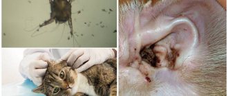
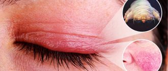
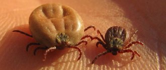
Why are ear mites dangerous in dogs? How to get rid of ear mites in dogs?
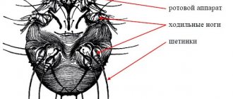
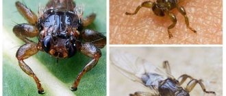
Ear mites in dogs and cats can live in any part of the ear canal, but they can only develop, multiply and parasitize in the ear canal. The ear mite feeds on lymph, which emerges when the parasite makes numerous punctures in the skin. This causes noticeable redness and irritation of the skin. In addition, the ear mite has a rough body, so when moving along the auricle and ear canals, the parasite severely injures the epidermis, which causes severe itching and burning. This causes severe discomfort to the animal, which affects its behavior. The pet begins to move a lot, scratches behind the ears, and shakes its head.
It is extremely important to promptly show your pet to a veterinarian and begin treatment, because ear mites reproduce very quickly: the female lays eggs, which after 4 days turn into self-feeding larvae. As a result of the life activity of the ear mite, numerous punctures become infected, and infections are added to otodectosis, otitis media and other diseases develop. The presence of even a few mites can cause otitis media in dogs and cats.
In advanced cases, otocdectosis can lead to inflammation of the eardrums, then the meninges, and even death.
See the smallest details
An endodontic microscope completely solves the problem of insufficient lighting and provides maximum detail of anatomical structures and/or materials previously used in treatment. By adjusting the degree of magnification, focus, and direction of the light beam, the doctor gets the most complete idea of the degree of development of the pathology, determines the location and scale of the lesion.
In addition, during the treatment process, photo or video recording of the visual field is carried out in parallel. In clinically complex cases, the doctor has the opportunity to review the examination record and consult with colleagues to develop optimal treatment tactics. The same recording will help to clearly explain to the patient the essence of the problem, the sequence and purpose of further medical manipulations.
Treatment of otodectosis in cats and dogs. Why do ear mites appear in dogs?
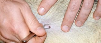
Only a veterinarian should prescribe a drug to treat ear mites. After all, you first need to find out at what stage of development the disease is, and whether there are any concomitant infections. If the presence of an ear mite is suspected, scraping of the contents of the ear canal from the inner surface of the ear is prescribed. Based on the results of tests and interviews with the animal owner, the veterinarian determines an accurate diagnosis and prescribes effective treatment.
As a rule, otodectosis is easily treated with anti-tick remedies in the form of ear drops or ointments for external use. If infections are observed to join the underlying disease, additional antibiotics and other drugs may be prescribed. The treatment regimen is completely individual and depends on the degree of development of the disease. If ear mites are detected early, the disease can be treated within a few days.
So, a dental microscope makes it possible to:
- conduct high-quality diagnostics of any dental disease;
- ensure super-precise fit of restorations;
- conduct microinvasive treatment;
- accurately determine the number, location and branching of channels;
- identify the features of the atypical structure of the canals;
- determine the degree of cleaning of the canal cavity;
- targeted action in the treatment of cysts and granulomas;
- remove fragments of instruments from tooth canals in the most gentle way;
- remove worn filling material;
- identify microcracks, perforations, sources of secondary infection and eliminate them.
Diagnostic methods
A patient entering for treatment at Nadezhda-Med LLC undergoes 4 types of diagnostic studies: Survey, Targeted, Special, Psychological.
Review diagnostics
Assess the general state of health in all organs and systems. And what is extremely important, they allow you not to miss any serious diseases that do not yet provide a clinical picture. For these purposes we use:
- “RUNO” diagnostics , which allows you to reliably see functional disorders (emerging diseases)
- "AMP" diagnostics - a high-precision diagnostic complex (130 tests in 1 hour, bloodless!) Diagnostics "AMP" - more accurate than conventional laboratory (with blood sampling) tests. We conducted special scientific comparative studies (click on the website menu “Books and Articles by A.G. Krivtsov” and read the article “Assessing the reliability and effectiveness of modern non-invasive diagnostic methods”
- More details: about RUNO diagnostics
- and AMP diagnostics (click).
MORE other ADVANTAGES of these diagnostics:
- bloodless, painless, non-invasive (does not damage the skin)
- fast (in 1 hour - !)
- provide the doctor with a huge amount of information (130 tests!)
- very inexpensive because it is a hardware complex, not separate tests. Both diagnostics are certified in the Russian Federation, as well as in a number of other countries - England, France, Germany, the United Arab Emirates, China, Ukraine, Kazakhstan, Belarus, etc. -cm. certificates. Diagnostics "AMP" has a certificate of the European Community (!).
We have been successfully using the “RUNO” diagnostics since 2000, and the “AMP” diagnostics since 2010. The simultaneous use of these 2 diagnostics in one patient means even greater reliability of the results.
“Targeted” (organ) studies
We prescribe only when we are absolutely sure of their need and only for any 1-2 or 3-4 organs. For example, we do NOT prescribe an ultrasound of all abdominal organs to a patient, but only an ultrasound of the liver and gall bladder. This allows us to reduce costs patient for very expensive hardware tests. The tests we most often prescribe are : ultrasound, MRI, ECG, EEG, REG, CT.
Ultrasound of internal organs
Ultrasound examinations in our Center are carried out only on high-quality equipment: - a new expert scanner - ESAOTE My Lab Class C (Italy) .
Special studies
We prescribe special studies - genetic or microbial flora, etc. only strictly according to indications. We refer patients to the appropriate laboratories.
Psychological research
Psychological research is a mandatory tool in the work of a psychologist and psychotherapist. There are also survey studies (for example, on the general condition of the nervous system) and targeted studies (for example, on the degree of depression, phobias, etc.). 600 psychological tests in our arsenal In addition, Professor A. G. Krivtsov , a doctor with 40 years of experience, a psychiatrist-narcologist, and a psychotherapist of the highest category, developed the following proprietary diagnostics to determine :
- severity (stages) of alcoholism;
- the degree of physical and psychological dependence from smoking;
- degree of obesity - based on a set of studies (12 parameters of body structure, taste preferences, level of metabolic processes);
- “ Narcological mirror” - determination of the level of pathological alcoholic stereotypes of thinking, which makes it possible to predict the outcome of treatment with a high degree of probability.
Any patient admitted to the clinic undergoes several stages of diagnostic tests and consultations.:
- initial consultation with a doctor,
- diagnostic tests before treatment,
- consultation of specialists,
- additional studies after consultation with specialists and during treatment,
- control studies for 1 year (1-3 diagnostics), in order to prevent a “breakdown” and prevent another exacerbation.
| Sign up for treatment | Sign up for diagnostics | Sign up for a consultation |
Eliminate with pinpoint precision!
By accurately determining the boundaries of the lesion, the doctor has the technical ability to act with pinpoint precision, preserving as much healthy tooth tissue as possible. This is a real chance to save a tooth, which just a few years ago would most likely have been considered hopeless.
High precision of teeth processing in preparation for the installation of veneers, lumineers and other structures for aesthetic restoration ensures their perfect fit to the surface. This is the key to achieving long-term results.
All medical procedures performed using a dental microscope are performed with the greatest possible comfort for the patient.
Working with an endodontic microscope is a high-tech type of medical care. This means that all our doctors have undergone additional training and are highly qualified.
The price of dental treatment under a microscope is higher than similar procedures performed using more conventional methods. The effectiveness, comfort and long-lasting results of treatment are truly worth the money.
How can I determine if my pet has otodectosis?
It is very difficult to see the tick itself with the naked eye due to its small size. However, it is quite easy to suspect the presence of these parasites in a pet. Urgently contact a veterinary dermatologist if your pet has at least one of the signs of otodectosis:
- The animal constantly scratches its ears;
- Irritation and redness appeared on the inner surface of the ears;
- The animal behaves restlessly;
- The appearance of discharge and unpleasant odor from the ear;
- The animal often tilts its head to the side.
Work examples
During routine sanitation of the oral cavity, a computed tomogram revealed that the root canals in the 36th tooth were not filled to the tops, but in fragments. Repeated root canal treatment was performed using a Carl Zeiss operating microscope.
The patient came to the clinic with pain when biting on the 36th tooth. A computed tomogram revealed an inflammatory process (cyst) in the area of the root apex. The anchor pin was removed and the root canals were re-treated using a Carl Zeiss operating microscope. View the work of specialists Before and After.
How to protect dogs and cats from ear mites? How to treat otodectosis in dogs?
The best way to protect your pet from otodectosis is to show attention to the animal:
- It is advisable to inspect and clean the animal’s ears after each walk, but at least several times a week.
- Closely monitor the behavior of your dog or cat.
- If you notice any discharge from your ears, contact your veterinarian immediately.
- Use antiparasitic drugs (shampoos, collars, sprays, etc.) only after consultation with your veterinarian. These drugs are very toxic and can cause allergies.
- If possible, avoid contact with stray animals.

