What does a taiga tick look like?
Judging by the finds of ancient ticks, their appearance has hardly changed over 380 million years.
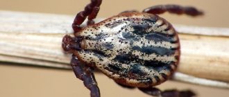
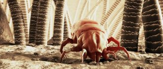
The body of taiga ticks is fused, usually oval in shape, pointed in front. The mouthparts of the taiga tick are located at the anterior end of the body or on its anterior-inferior side. On the back of ticks there is a shield that occupies the entire surface of the body in males, and about a third of the body in females. The streamlined shape facilitates movement both in the forest floor substrate and on the host’s body. Hungry adults have a length of 2-6 mm, satiated ones - 10-20 mm. Their integuments are sometimes hard, inextensible, partially soft and elastic, collected in folds, which allows them to absorb large amounts of blood. In the photo you can see a taiga tick that has already had its fill. The color of sucked parasites varies from dark gray to reddish, and their weight increases a hundred times or more.
Ixodid ticks. External structure and oral apparatus
Sem. Ixodidae - ixodid ticks have 713 species. It is accepted to divide these mites into 2 groups: Prostriata (Ixodinae) and Metastriata (Amblyomminae) with one or more subfamilies. In the latter case, to the subfamily.
External structure and oral apparatus of ixodid ticks. Medium and large ticks. The body length in hungry females of the genera Amblyomma and Hyalomma reaches 0.7-10 mm, and in fed females - 20-30 mm with a weight of 1000-2000 mg. In smaller species from other genera, the body length of engorged females does not exceed 5-15 mm. The body size of the larvae is less than 1 mm. The anterior half of the dorsal surface is the idiosome, and in males the entire surface is occupied by the dorsal shield, or scutum.
The integument of the idiosome, except for the dorsal scute, is assembled into a system of epicuticular parallel microfolds, the straightening of which, together with the growth and stretching of the underlying procuticle, ensures a multiple increase in body volume during feeding. In the adult phase, sexual dimorphism is pronounced, manifested in the preservation of greater sclerotization of the idiosome in males, in the structure of the gnathosoma and genital openings. Larvae and nymphs are more similar in appearance to females, but differ from adults in the structure of the gnathosoma, sensory apparatus, chaetotaxy and the absence of genital openings. On the head there is a terminal proboscis, forming a cutting-sucking type mouthparts. The lower half of the proboscis is formed by an extension of the base of the gnathosoma, the hypostome, extended forward. The ventral surface is convex and armed with parallel rows of posteriorly directed teeth. The dorsal half of the proboscis is formed by tubular cheliceral cases, which represent the anterior outgrowth of the roof of the base of the gnathosoma.
Inside the cases are long rod-shaped chelicerae, consisting of a long shaft and a finger. The cheliceral trunks are capable of moving in the anteroposterior direction inside their cases. Movable fingers of the chelicerae can make cutting movements in the mediolateral plane, due to which cutting through the skin of vertebrates occurs. A tubular preoral cavity is formed between the dorsal surface of the hypostome and the ventral cheliceral sheath. In the posterior part of the proboscis, the hypostomal groove deepens, and the hypostomal membrane closes over it on the dorsal side, thus forming a mouth that leads directly into the pharynx and has a triangular cross-section.
Taiga tick: photo
In order to make sure that this is a taiga tick, look at a photo of the pest and compare the key external characteristics of the parasite with the structural features of the individual you discovered.
- The size of an adult taiga tick varies from 2 to 6 millimeters, not counting the legs.
- The body of a hungry pest is flat; the tick and head are teardrop-shaped.
- The body is colored brown and dark brown, which are superimposed on each other in the form of two circles of different diameters.
- Females are usually larger than males.
- Once engorged with blood, the tick swells up to 1.5 centimeters in diameter and takes on the three-dimensional shape of a bean.
- The color of a well-fed tick becomes gray due to the blood filling its body.
- The insect has 8 legs - 2 pairs of long ones in front and behind and 2 pairs of short ones on the sides.
Ticks do not move particularly quickly, so if the insect has not yet had time to attach itself to you, it will be easy to remove it. Place the pest in a jar, bottle or any other transparent container using tweezers, a piece of cloth, a cotton pad or something else, the main thing is not to do this with your bare hands. If the size of the tick is too small, take a good photo of it and zoom in on the photo on your phone screen. This will give you the opportunity to examine the parasite in detail without risking your own health.
PLIERS
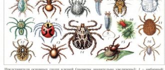
Representatives of the main groups of ticks (sizes are significantly increased): 1 – granary; 2, 3 – armored; 4 – iron; 5 – feather; 6 – itching; 7, 8 – press...
Ticks (Acari, Acarina), a group of chelicerates of the arachnid class. The question of the unity and rank of the group remains debatable: they are considered either as a single subclass or class of animals, or as a combined group, including the orders of Acariformes and Parasitiformes. order or classified as parasitiform K. Fossil remains of K. are known from Devonian deposits. By origin, K. is associated with trilobites. Body length is from 80 µm to 13 mm, in engorged bloodsuckers it is 30 mm. The body is unsegmented or divided in acariform K. into a proterosome (segments bearing chelicerae and pedipalps, the first and second pairs of legs) and a hysterosome (segments with the third and fourth pairs of legs and abdomen), in parasitiform K. - into a prosoma (cephalothorax) and opisthosoma (abdomen). In primitive forms the body bears traces of segmentation. The integument ranges from weakly sclerotized with compacted areas in the form of shields to a dense cuticular carapace, color from whitish-yellow to bright red or dark brown, almost black. The oral apparatus is mainly gnawing and piercing-sucking type. In most species of K., the chelicerae are claw-shaped with pronounced teeth; in spetsializir. species can be modified; pedipalps 3–6-segmented. The bases of the pedipalps merge to form the preoral cavity.
Some species of K. have eyes. Breathing is cutaneous or tracheal. The development cycle from egg to mature individual includes a prelarva (typical of acariform mites and harvest mites; inactive, does not feed, can develop hidden under the protection of egg membranes and be reduced to one degree or another), a larva and 1–7 nymphal stages. Prelarvae and larvae of K. have 3 pairs of legs, nymphs and adults have 4 (the exception is gall mites with 2 pairs of legs). Most K. have pronounced sexual dimorphism. The methods of fertilization in K. are varied: they deposit spermatophores on a substrate (indirect fertilization) or transfer spermatophores or sperm to the female during copulation (direct fertilization); Parthenogenesis is often observed. Fertility - from several tens to 20 thousand or more eggs. The development time of one generation ranges from several days to several years.
OK. 50,000 species. K. are found on all continents, inhabit the soil, various. organic remains, nests and burrows of animals, plants, decomposition. reservoirs, etc. The number of soil mites can reach 500 thousand individuals per 1 m2 of soil, parasitic ones - more than 10 thousand per animal (red mite larvae). The trophic specialization of K. is diverse: among them there are saprophages, predators, phytophages, obligate and facultative hematophages (blood-sucking), etc. Many K. are characterized by passive dispersal with the help of other animals - phoresy. Some species of K. associated with animals can switch to ecto- and endoparasitism.
K. parasitic on the body of humans and animals cause dermatitis, scabies and other diseases; House dust mites and barn mites are sources of allergens. K. transmit more infectious agents. diseases than any other group of arthropods. They serve as carriers of viruses, bacteria, protozoa and filariae - pathogens of various types. diseases of humans and animals (tick-borne encephalitis, babesiosis, theileriosis, tularemia, etc.). In natural foci of vector-borne diseases, ixodid ticks are the most dangerous. Some species of oribatid mites serve as intermediate hosts of helminths that parasitize agricultural crops. animals. Mn. K. species damage cultivated plants (spider mites, gall mites, etc.), stocks of grain, flour and other food products, and damage plant bulbs and tubers during storage (barn mites). A number of K. species carry pathogens of plant diseases. The Varroa destructor mite causes significant damage to beekeeping. At the same time, armored mites living in the soil play an important role in the cycle of substances and soil formation, and predatory gamasid mites are used to control plant pests (see Biological method of plant protection). The science of cancer is acarology.
Appearance of the taiga tick
Heterogeneous individuals of the taiga tick are similar in structure. Both have a proboscis, head, torso and legs, but they look different.
Females in a hungry state are red-brown in color, 3-4 mm long. Having drunk blood, they increase in volume by 12-13 times, and their weight can increase by 100 times. Special folds allow the body to expand. A well-fed female changes color and becomes gray, acquiring a swollen oval shape. A third of the body is covered with a black chitinous shield.
The male is smaller, 2-3 mm long and the shield covers the body completely, so it looks black. Males do not increase in size, since they do not need to feed for a long time and a lot.
How to distinguish a taiga tick from an insect or spider? The morphology of ticks reveals several differences in the structure of the body:
- almost all mites have eight legs, while insects have six;
- the presence of a mouthpart (gnathosoma) distinguishes ticks from spiders or scorpions. The taiga tick has a piercing-sucking mouthparts;
- lack of eyes.
Mouthparts of a tick | Your home dermatologist
The structure of the oral apparatus was studied on serial sections of females of Ornithodoros papillipes and Hyalomma asiaticum and sections of the host integument together with the H. asiaticum nymphs attached to them in varying degrees of saturation, as well as on whole preparations of individual dissected parts of the gnathosoma. The anatomical structure of the gnathosoma was discussed earlier, and this section discusses its functional and morphological features necessary for understanding the mechanism of food intake.
Hyalomma asiaticum. The lower half of the proboscis is formed by an extension of the base of the gnathosoma, the hypostome, extended forward. The latter is slightly spoon-shaped, widening and flattening towards the distal end. Its ventral surface is convex and covered with rows of powerful teeth bent back. The dorsal surface of the hypostome is slightly concave and bears a food groove medially. In the distal part it is represented by a relatively shallow U-shaped depression. Proximally, the groove deepens, is flanked on the sides by two longitudinal ridges of the epicuticle, and its lumen in a transverse section becomes V-shaped. In the distal part of the food furrow, the cuticular layer is tightly adjacent to the exocuticle, and in the proximal part it moves away from it and forms the so-called hypostomal membrane, so that free space is created between them.
In the works of R. Arthur, this hypostomal membrane is mistakenly interpreted as an epipharynx, fused at the edges with the hypostome, and the cavity covered by it from above as a food canal. Our observations force us to abandon such an interpretation, since the food canal passes not under, but above the hypostomal membrane, and the cavity located under it is absent in the anterior half of the proboscis, not opening outward. D. Gregson shares a similar opinion. In Hyalomma asiaticum and other species we studied, the rise and fall of the membrane turned out to be very insignificant, purely passive and unlikely to significantly affect changes in the volume of the preoral cavity during feeding.
Inside the hypostome, along its entire length, a dorso-ventral suture can be traced, indicating the primary paired origin of the hypostome as a result of the fusion of the anterior processes of the coxal pedipalps. There is a channel in each half. Both canals merge in the proximal part of the hypostome into one unpaired one, representing a continuation of the body cavity. In recently molted individuals, the hypodermal lining is still preserved in these internal cavities.
In the ticks Ixodes ricinus, I. hexagonus, and I. persulcatus, groups of dorsoventral muscles are described in the hypostomal cavity, extending from the lateral walls of the food furrow to the ventrolateral corners of the cavity. In a detailed study of complete series of sections, we found such muscles only in the most proximal region of the hypostome of I. persulcatus, but they were absent in Hyalomma asiaticum, Dermacentor pictus and Rhipicephalus turanicus. Apparently, hypostomal muscles are characteristic only of species of the genus Ixodes, and in this respect they are similar to argasid mites.
The dorsal half of the proboscis is formed by chelicerae. The cheliceral trunks are surrounded by double tubular cuticular sheaths, the origin and structure of which is still controversial. The most powerful outer cases undoubtedly represent the anterior outgrowth of the roof of the gnathosoma. It splits at the anterior end and reaches dorsally with retracted chelicerae to the bases of the fingers. On the ventral side they are somewhat shorter and thinner, so that in the anterior part of the proboscis on transverse sections they initially look like the roof of trunks. Curving ventrally, they enclose the latter in hard tubes. The comb-like mediodorsal outgrowths of the walls of the trunks enter the corresponding grooves of the cases and prevent unwanted lateral movements when the chelicerae slide in the anteroposterior direction. In the distal part of the proboscis, the outer cheliceral cases are independent, but tightly adjacent to each other with almost straight inner walls. Before reaching the base of the head, they merge first along the dorsal and later along the ventral border, so that a solid semiring is formed, separated by a vertical septum. The lower wall of the outer cheliceral cases continues into the subcheliceral plate embedded in the base of the gnathosoma.
In the distal part, the outer dorsal surface of the cheliceral cases is covered with numerous small denticles, facilitating fixation on the host during feeding. The distal ends of the outer cheliceral cases are wrapped inward and, in the form of a membrane-like shell, go backward, attaching to the cheliceral trunks in front of the pharyngeal opening. Similar formations are described in Dermacentor andersoni and are called conical cheliceral sheaths. Apparently, they play an essential role in the protrusion of the chelicerae, preventing the penetration of blood and foreign particles into the cheliceral cavity during feeding.
Behind the conical cases, the cheliceral trunks are again surrounded by a membranous membrane extending dorsally from the inner surface of the tectum, and ventrolaterally from the subcheliceral plate. The morphological interpretation of these formations is difficult, and they are usually called internal cheliceral sheaths.
The anterior part of the base of the gnathosoma is characterized by the greatest complexity of structure. The fusion of the outer cheliceral cases and the hypostome into a dense chitinous ring occurs at a considerable distance in front of the oral opening, so that an extensive morphologically intact tube of the preoral cavity is formed, 0.32 mm wide and 0.04–0.08 mm high. Its roof is the ventral wall of the outer cheliceral cases, and its bottom is the dorsal surface of the hypostome; the wall and surface are connected to each other by lateral-ventral processes of the coxal palps. In the anterior third of the proboscis, there is no morphological fusion between its components, but due to their close fit to each other, the preoral cavity actually continues to the distal end of the hypostome.
A food groove runs along the midline of the dorsal wall of the hypostome. In the distal part it is relatively shallow, and in the proximal direction it gradually deepens to 0.04 - 0.06 mm and acquires a V-shaped cross section. The oral opening is located in the depths of the preoral cavity in the place where the food groove is covered from above by a short cone-shaped labrum.
The portion of the preoral cavity that extends posteriorly over the labrum and the dorsal surface of the subcheliceral plate is called the salivary reservoir. It is strongly flattened dorsoventrally, and the main excretory ducts of the paired salivary glands, measuring 0.07×0.03 mm, open into its posterolateral corners. The latter first run in the thickness of the subcheliceral plate, and then separate from it in the ventral direction. The ceiling of the salivary reservoir is formed by the ventral wall of the outer cheliceral sheaths, which passes into the subcheliceral plate at the junction of the salivary ducts.
The subcheliceral plate serves as the main supporting structure for the chelicerae and pharynx. In the anterior part, it is connected by ventrolateral processes to the walls of the base of the gnathosoma and forms an intercoxal bridge. Further back, it loses connection with the walls of the gnathosoma and becomes greatly thickened. After merging with the ventral wall of the outer cheliceral sheaths, a high vertical ridge runs along its dorsal surface. It represents a continuation of the fused medial walls of the outer cheliceral sheaths and passes dorsally into the inner cheliceral sheaths. On the ventral side, muscle bundles coming from the pterygoid processes and dorsal pharyngeal dilators are attached to the thickened edges of the subcheliceral plate. The anterior end of the subcheliceral plate continues into a short labral process.
Ornithodoros papillipes. The structure of the oral apparatus of the species in question, according to the general structural plan, is very similar to that of ixodid ticks, but there are a number of differences associated with different methods of blood absorption in both groups. The proboscis of Argasid mites is thinner and is not adapted for long-term attachment of the tick to the host's body, while the fingers of the chelicerae, on the contrary, are more powerful and are adapted for quickly cutting through the integument.
The degree of development of the hypostome teeth, the size of the movable fingers of the chelicerae, and the terminal or ventral position of the gnathosoma are largely related to the duration of feeding. The hypostome, armed with regular rows of teeth directed backwards, serves as the main organ of anchorage in the host’s integument. The longer the blood-sucking continues, the more firmly the tick should be held on the host’s body and the more developed the hypostome armament should be.
For fast-feeding species, consolidation is not so important and the priority is to absorb the full portion of blood as quickly as possible. Therefore, the degree of development of the cheliceral fingers, which ensure penetration into the integument of the host, plays a significant role for them. Inside the proboscis, just like in ixodid ticks, there is a tube-shaped preoral cavity. Transverse sections show that it is compressed in the dorsoventral direction into a flattened canal. In the distal part, this canal is limited by closely adjacent bent edges of the chelicerae and a slightly concave flattened hypostome. At the level of the beginning of the cheliceral sheaths, the tubular canal is preserved due to the corresponding bends of the ventral surface of the outer sheaths and the increasing concavity of the hypostomal groove along with its marginal processes. Further back, unlike ixodids, it is divided by an elongated upper lip - a “stylet” into dorsal and ventral halves, representing independent canals.
The lower half of the preoral cavity is limited below and laterally by the bottom of the medial hypostomal groove, which gradually turns into a deeper canal. From above it is covered with the upper lip. The dorsal surface of the latter, together with the lateral zones of the dorsal wall of the hypostome, form the bottom of the upper half of the preoral cavity. The ceiling of the latter is formed by the ventrally fused outer sheaths of the chelicerae. The lower canal ends in an almost triangular oral opening leading to the pharynx. The superior tube opens into a highly flattened blind sac of the salivary reservoir. Its lower wall is the epistomal plate, and its upper wall is the ventral surface of the outer sheath of the chelicerae. The ducts of the salivary glands open into the posterolateral corners of the described cavity. In the literature it is unreasonably called buccal, i.e. oral, although it has nothing to do with the oral opening. Functionally, it represents a reservoir in which saliva flowing from the ducts of the salivary glands accumulates.
Evolution of the oral apparatus. Among parasitic ticks, the oral apparatus of ixodid ticks represents the extreme degree of specialization for feeding on blood. Nevertheless, it is possible to identify the main structural components characteristic of other arachnids. A detailed morphological analysis of the oral apparatus of arachnids performed by R. Snodgrass allowed him to recreate the original prototype common to all orders.
In adults, the part of the prosoma derived from the acron includes the labrum, the epistomal region, and a narrow medial strip of the anterior body wall connecting the epistome with the carapace between the bases of the chelicerae. The somites of the chelicerae remain postorally, but the chelicerae themselves, as a result of preoral movement, occupy an anterior position, and their sternal areas are almost completely reduced. The pedipalps, excluding Palpigradi, also move forward on the sides of the mouth and connect dorsally with the epistome, forming the intercoxal bridge. The sternal plate of the pedipalpal segment forms the deutosternum, which projects in front of the oral opening and forms the lower lip.
The mouth is immersed in the preoral cavity, bounded laterally by the coxae of the pedipalps, below by the growths of the coxae of the pedipalps or deutosternum, and above by the labrum, and leads into the pharynx of the sucking type. The oral apparatus of ticks has undergone significant modifications in comparison with the above diagram, which in general terms boil down to the following. Chelicerae and pedipalps are united in an independent part of the body - the gnathosoma. It also includes most of the preserved acron, while the ocular region of the latter, like other arachnids, is significantly displaced back and merges with the tergites of the prosomal segments. The dorsal surface of the gnathosoma is sclerotized over the bases of the chelicerae and forms a roof, or tectum. Laterally, the latter merges with the dorsal growths of the gnathocox pedipalps, which simultaneously merge with each other ventrally along the midline. As a result, a hard chitinous ring at the base of the gnathosoma is formed, characteristic of most Parasitiformes, and according to Snodgrass in general of all mites.
Ventrally, the anterior coxal processes of the pedipalps extend forward, merge along the midline and form a hypostome, representing the floor of the preoral cavity. The oral opening is covered from above by a labrum, which is attached at its posterior end to the subcheliceral plate. The latter is homologous to the epistome of other arachnids, since it always participates in the formation of the intercoxal bridge, and dorsal pharyngeal dilators are attached to its lower surface. Embryological observations of gnathosoma formation largely support Snodgrass's main points.
The closest thing to the hypothetical prototype of the oral apparatus of ticks is the gnathosoma of some free-living gamasids, such as Poecilochirus carabi, studied in detail by W. Winkler. Complications in its structure are associated with the appearance of membrane-like cheliceral sheaths, with various modifications of the hypopharynx and labral processes, as well as with the development of paired anterior hypostomal processes.
Recently, A. B. Lange, relying on the views of A. A. Zakhvatkin on the polyphyletic origin of ticks, put forward a new interpretation of the segmental composition of their oral apparatus. A. B. Lange believes that the gnathosoma in different orders of mites includes a different number of somites, and there is no clear intersegmental boundary between this section and the prosoma. In this regard, he suggests using a more neutral term - gnatema. Without touching on the highly controversial issue of the phylogenetic relationships of different groups of mites, it seems to us that in relation to the order or suborder Parasitiformes there is no reason to abandon the generally accepted term gnathosoma, regardless of the systematic rank of this group.
Its morphological composition is uniform in all Parasitiformes, and the absence of an intersegmental boundary with the prosoma is not of fundamental importance. The posterior border of the prosoma with the opisthosoma in many arachnids is also not intersegmental, but this does not raise doubts about the independence of these tagmata. The transition to feeding on blood introduced significant changes in the structure of the gnathosoma of ticks, and the exclusively specialized oral apparatus of ixodids can only be understood when compared with that of the more primitive blood-sucking gamasids, which have been studied in detail in this regard. The oral apparatus of Haemagamasus nidi is characterized by the preservation of many primitive features, in particular massive pincer-shaped chelicerae with rough teeth on the edges of both fingers, as well as a wide and short oral cone with an open pre-oral cavity. This species is not yet capable of receiving blood by actively puncturing the host’s skin, and is characterized by a mixed type of nutrition, including eating dried or semi-liquid blood around wounds, schizophagy and entomophagy.
Dermanyssus gallinae, Sauronyssus saurarum and Ornithonyssus bacoti are representatives of the group of obligate bloodsuckers. Their chelicerae are transformed from gnawing organs into piercing-cutting organs. Their trunks become thinner and significantly lengthen. At the same time, the oral cone is stretched and a tube-like preoral cavity, almost closed from the sides, is formed. These species already independently cut through the integument of vertebrates and drink the blood flowing from the wounds. Ticks over everything. Ixodoidea are among the most advanced bloodsuckers and their oral apparatus is adapted to absorb blood directly into the thickness of the host’s skin, where it immerses while feeding. The long rod-shaped chelicerae have completely lost their primary claw-like shape, and the number of their segments is reduced to two. The mobility of their cutting fingers is limited to the mediolateral plane, and they have evolved into an extremely efficient cutting organ, easily penetrating the hard stratum corneum of the epidermis.
The outer cheliceral cases and the hypostome are closely adjacent to each other on the sides and form a proboscis, strongly elongated in the anterior direction, with an internal tube-like preoral cavity. The latter serves as a conductive channel that pumps blood or infiltrate directly from the inner layers of the dermis, which are very rich in blood vessels.
Along with these common features, significant differences in the structure of the gnathosoma are observed between ixodid and argasid ticks. They are predominantly adaptive in nature, associated with various types of parasitism in these families; they are discussed in detail in the previous section. According to a number of morphological characteristics, the oral apparatus of the argasid is less specialized compared to ixodid ticks. Their proboscis is less elongated and a long epistome is preserved inside it, dividing the preoral cavity over a considerable length into the ventral food and dorsal salivary canals. All four palpal segments are well developed and movably connected to each other.
‹ Adaptation to increase in tick body size during feeding Up Structure of salivary glands in ticks ›
Lifestyle of the ixodid tick
Everyone should familiarize themselves with information about the lifestyle of this arachnid. Knowing where these parasites live, how they reproduce and what they eat will help you minimize the likelihood of encountering them when going outdoors. Such information will help not only to avoid an attack by the parasite, but also to prevent the development of infections carried by the taiga tick.
Habitat, conditions suitable for life
In Russia, ixodes persulcatus is found mainly in the middle and southern subzones of the taiga. The habitat of arachnids extends to the west, covering the Moscow and Leningrad regions, and in the north – the southern regions of Karelia. In the Volga region, the parasite lives in the Ulyanovsk and Samara regions north of the Samara River.
Ixodid ticks are found in Belarus, the Baltic states, on the southeastern coast of Finland, and in certain parts of northwestern Europe. To the east, their habitat extends to the Pacific coast. The parasite also lives in the southern regions of Kamchatka and Japan, on Sakhalin, the Kuril Islands, and in isolated areas of the mountain ranges of Central Asia.
Moisture is important for the comfortable existence of the taiga tick, which is why it is an inhabitant of forest habitats. It can also be found in meadows, ravines and ravines with tall, dense vegetation and bush thickets.
Recently, parasites have begun to appear in green areas of the city, in summer cottages located in the suburbs, cemeteries, parks, and recreational areas of populated areas, where they migrate on birds, rodents and other animals. They are active at temperatures above 0.3°C. At temperatures above 20°C they become less active.
Reproduction and development stages
The life cycle of the parasite follows a pattern of complete transformation. Its ontogenesis includes 4 morphological stages: egg and 3 active phases, separated by molts. The penultimate 2 stages of development are immature (preimaginal). These are the larval and nymph stages. The last phase of metamorphosis is sexually mature, i.e. imaginal.
The process of active mating begins at the end of spring, when a stable above-zero temperature is established. Parasites mate both in their natural habitat and on the body of the victim. Before laying eggs, the female feeds heavily for at least 7 days. Masonry occurs on soil and plants. The number of eggs laid by one female can reach several thousand. At the same time, only a few adults survive, the rest die due to unfavorable conditions or hunger.
Several weeks pass before the taiga tick larvae appear. At this stage of development, the size of future nymphs barely reaches 0.9 mm. The larvae have only 3 pairs of legs. For several days they feed on the blood of animals or birds. Having thoroughly sated, the larva leaves the victim’s body, returning to its natural habitat. After molting, a nymph is born with a full set of limbs.
In this phase of ontogenesis, the parasite waits out the winter in order to continue its transformation with the onset of warmth. The nymph turns into an imago no earlier than a year after the last molt. Characteristics of a tick at different stages of metamorphosis:
Parasite nutrition
These arthropods are polyphagous, so their food supply is extensive. The source of food for bloodsuckers are various species of mammals, birds, reptiles, ungulates, predatory animals, and pets (dogs, cats). In the preimaginal phase, individuals feed on the blood of mouse-like rodents, chipmunks, squirrels, hares, insectivorous birds, etc.
While waiting for a victim, the parasites climb to a height of no more than 1 m. They can remain in this position for up to 4 weeks. Sensing the approach of a food source, the tick puts forward its two front pairs of limbs, clinging to the victim’s body with them. Finding a suitable area of skin, it attaches itself and pierces the skin. At the site of the bite, blood components begin to decompose, and the parasite absorbs it in a half-digested form.
Humans are usually hunted by adults, and extremely rarely by nymphs. These arthropods do not have eyes. The lack of the ability to see is compensated by excellently functioning olfactory organs and thermoreceptors. Parasites are able to react to the heat and smell of a victim’s sweat at a distance of up to 10 m. Without food, they can exist from 1 month to 3 years.
Natural enemies
These arachnids have enemies in nature, the main of which are the ichneumon wasps Ixodiphagus hookeri and Ixodiphagus hirtus. In our country, they are found in Uzbekistan and the Far East. The females of these insects lay eggs in the body of the female parasites. The larvae that hatch from them feed on the insides of the host tick, leaving only a chitinous cover. Ixodes persulcatus serve as food for ground beetles, ants, birds, in particular thrushes, spiders, frogs, and lizards. Another enemy of parasites is parasitic fungi, which massively destroy their nymphs.
Habitat
The taiga tick lives over a vast territory and is found on almost the entire Eurasian continent. Representatives of this species can be found in Altai, on the territory of the Kuril Islands, in the central part of the country and, of course, in the west.
The bulk of the population lives in the taiga, which is how the species got its name . The insect prefers deciduous forests and can be found in forest-steppe.
As you move south and humidity levels drop, the number of ticks decreases. Far from civilization, where there are no people and no cattle, the parasite can be found in the burrows of warm-blooded animals.
What are the differences between the taiga tick and others?
In appearance, the taiga tick is similar to many other species of ixodid ticks belonging to the arachnid class. But there are also differences in the structure of the body: a single-color black-brown dorsal shield, an anal groove that goes around the anus in front and the absence of eyes. The European tick differs from the taiga tick in the presence of small membranous appendages and long internal spines along the posterior edge of the coxa I, as well as the straight posterior edge of the base of the proboscis on the dorsal side. The forest tick is characterized by two increases in numbers, in contrast to the taiga tick.
The taiga tick differs from the dog tick in some structural features and a shorter life cycle. Depending on the conditions, the development of the taiga tick can continue for 2 or 3 years with a change in several hosts, and the full cycle for the dog tick is 3-4 months.
Interesting! A biological feature of taiga ticks is negative geotaxis, which is expressed in the desire of parasites, after attaching to a host, to crawl upward, against the gravity of the Earth, deviating only slightly to the sides.
Cycle of life
On average, the full life cycle of an insect takes about a year, but there are cases when it lasted up to 7 years in the absence of favorable living conditions.
The stages of development of the taiga tick include the following phases:
- egg. It has a yellowish or brown color, the size does not exceed 1 mm. The female lays eggs after receiving a portion of blood, usually in the spring. A clutch of 2–3 thousand eggs can be formed at a time. They all lie in a heap in the grass for 2-3 months, waiting to move on to the next stage of development.
- Larva of the taiga tick. Its shape resembles an adult insect, it is capable of moving short distances and attaching itself to the victim’s body for several hours. The body size of the taiga tick at this stage does not exceed 1 mm.
- Nymph. They are similar in appearance to an adult insect, but differ in size. The body length at this stage is about 1.7 mm. The nymph forms inside the larva, emerging after molting. Sticking to its victim, it remains on it for up to 8 days. In the nymph state, ticks can overwinter, falling into suspended animation and becoming more active as soon as the outside temperature reaches positive values.
- Adult insect. The insect reaches this stage of maturity after the nymph has moulted. An adult insect needs blood and immediately begins to look for a new victim.
The peak of parasite activity occurs in May–June, when hungry ticks crawl out of their shelters in search of food.
Having completed their reproductive task, most of the insects die and their total number decreases. Taiga parasites become active in the fall, when the first larvae hatch from the eggs.
Appearance of the parasite
The popular belief that a tick is an insect is completely wrong. In fact, this parasite is an arachnid.
The length of a tick is usually no more than three millimeters. The average length of the parasite is even less: 0.1-0.5 millimeters. Does a tick have wings? Just like any arachnid, the parasite has no wings at all. However, adult individuals have four legs, while forest ticks that have not reached puberty can boast only three.
It is interesting that the tick does not only have wings: the bloodsucker also does not have eyes. This does not prevent them from being able to navigate the terrain well, due to their very developed sensory apparatus. It is thanks to their developed senses that forest ticks can smell a possible victim at distances that are large for their size - ten meters.
Ticks can be divided into separate sections:
Leatherbacks with fused head and chest. They inhale air using the trachea and skin. Photos of the tick are presented below
Armored with a head attached to the body. Breathing in this species is possible with the help of special spiracles. Photos of the tick are presented below
Reproduction and life expectancy of the taiga tick
At the end of spring, taiga ticks undergo the mating process either in their natural habitat, or already on a host while the female is feeding. After complete saturation, the female lays 1.5–2.5 thousand eggs, from which larvae no larger than 0.5 mm in size and six legs will appear within a few weeks.
For further development, the larvae feed on the blood of small animals or birds for half a week and again return to their natural habitat, where they molt and turn into nymphs (i.e., they move to the next phase of maturation).
Ticks in this phase differ from previous ones in their larger sizes (up to 1.5 mm) and the presence of 8 legs. At this stage, they go to winter, after which they hunt again, and this time warm-blooded animals, including humans, become food objects for further development.
Then the nymphs again experience the process of molting, after which they turn into adults the next year. It follows that the lifespan of a taiga tick corresponds to the period of its full development and takes at least 3 years (although this process sometimes drags on for 4–5 years).
During this period, under the influence of various natural conditions and other factors, from a large number of larvae to the stage of an adult tick, only the strongest survive (only a few dozen).
To summarize, I would like to remind you once again that the taiga tick is the causative agent of the most dangerous diseases (and only adult individuals are dangerous to humans), therefore, when going to the forest in the summer, you need to follow the simplest rules to ensure protection from these creatures.
They consist of regular inspection of clothing, limiting sitting on the grass and moving in thickets, using repellents, and upon returning home, a complete change of clothing and a thorough examination of the body. Also, vaccination against encephalitis, which is constantly carried out in populated areas during the period of active “hunting” for ticks, would not be superfluous.
Reproduction
Regardless of the duration, the life cycle of all such animals is the same: the female lays eggs, which eventually transform into larvae. The latter, after a certain period, turn into nymphs - semi-adult individuals that do not have reproductive capabilities and have three pairs of legs. After passing through three stages (protonymph, deutonymph and tritonymph), they turn into adult ticks (imago). Males die over time, females lay eggs. The cycle repeats.
Interesting: Are there right-handed and left-handed animals?
There are varieties whose females do not need males (the so-called parthenogenetic method of reproduction). Their cells begin to divide under the influence of certain environmental factors. Other species reproduce in the usual way. In some species of ticks, females do not live to see the larvae, which, once born, independently gnaw their way out of the mother's womb.
What is the taiga tick transmitting?
The taiga tick is dangerous because it is a carrier of pathogens of many human infections, the most severe of which is tick-borne encephalitis. In addition, a tick bite can lead to other pathologies. At least 18 bacterial pathogens are known to be transmitted by ticks: 8 Rickettsia species, 3 Ehrlichia species, 4 Borrelia species, 3 Bartonella species. Diseases transmitted by ticks include borreliosis (Lyme disease), relapsing fever, tularemia and some other, rarer infections.
Danger to humans
Reports about the danger of taiga ticks are becoming more common every year. This is primarily due to the fact that populations of these parasites have become more numerous and the risk of being bitten has become more real. A tick bite is an opportunity to contract a dangerous disease that is transmitted through the parasite’s saliva.
Through the bite of a Siberian tick, a person can become infected with:
- encephalitis, which leads to damage to the nervous system, and in the case of an advanced stage of the disease, even death;
- borreliosis or Lyme disease, which affects the functioning of the cardiovascular and nervous systems, affects vision and joint tissue;
- babesiosis, which is characterized by fever, acute pain in the abdomen and muscles. Often this virus leads to the death of the patient, since the disease is difficult to recognize;
- ehrlichiosis, which causes destruction of the kidneys, liver and spleen. It can also cause pulmonary and gastric bleeding.
In each case of the disease from a tick bite, the patient requires constant medical supervision and hospital treatment. The ideal development of events is if the tick that has bitten a person is detected in time, removed and submitted for analysis to a special laboratory. This allows you to find out what viruses the insect carried and prevent the development of the disease in time.
Prevention of bites
In order to significantly protect yourself from tick bites and notice the danger in time, when going into nature, you should follow the rules:
- Wear clothes made of thick fabric, trying to cover as much of the body as possible.
- The collar of the jacket should be tied, and the pants should be tucked into boots.
- After returning home, it is better to inspect your outerwear and shake it outside.
- You also need to examine your body (for convenience, you can ask your loved ones about this).
- It is better not to move through dense thickets of grass or bushes unless necessary.
- Moreover, it is recommended not to sit or lie down in it to rest.
- If infection with tick-borne encephalitis is not uncommon in the region, then it is rational to get vaccinated in advance.
To protect your household, it is recommended to trim the grass around your home regularly. For pets that can also bring ticks, repellent sprays and collars are used.
Information about what the insect is and why the taiga tick is dangerous will be useful to everyone who lives in its habitat or is planning a trip to such a region. Having discovered a tick, you need to properly remove it and submit it for analysis to find out whether it was a carrier of infectious diseases.
Development and growth of taiga ticks
The taiga pest develops from eggs, which the adult female lays in huge quantities (up to 3 thousand at a time). The next stages of growth are the larva and then the nymph. During this period, the tick already leads an active lifestyle and feeds at the expense of the owner. Over time, the parasite grows to an adult state. The full development cycle of the taiga tick takes from two to six to seven years.
During its life, the taiga tick goes through 5 stages
How taiga ticks reproduce
The male waits for his mate in the grass or clinging to the owner. Having replenished its reserve of strength, it falls away from the victim and fertilizes a large number of females. After completing the feat, the male taiga tick dies (by June there are no adult males left, but larvae and nymphs go hunting). The fertilized female feeds from the owner for another week, after which she lays eggs. At this time, its size can reach 1 centimeter.
The female tick usually reaches a very large size because she eats more than usual while laying eggs.
Life cycle
The life cycle of the taiga tick follows a principle common to all ixodid ticks (see tick reproduction). Ontogenesis lasts 3 years. During this period, Ixodes persulcatus goes through 4 stages of development: egg, larva, nymph and adult (imago). The transition between these stages is molting.
Nutrition is very important for the full development of ticks. For the eggs to develop, the female needs to be fully nourished. Only individuals that do not need food also molt.
The taiga tick is also characterized by a change of hosts. Larvae and nymphs feed on small rodents and birds that lead a terrestrial lifestyle. Adults (imagoes) prefer large mammals, in particular humans.
Who does the taiga tick attack?
This bloodsucker by its nature is a polyphagous polyphagist, that is, it drinks any blood it comes across, without particularly sorting.
Victims of an attack by a taiga parasite can be:
- Small rodents;
- Predators;
- Large mammals;
- Birds;
- Domestic warm-blooded pets, especially dogs and cats;
- Livestock, horses.
- People.
In the larval and nymph stages, small individuals are accessible to the parasite; it also does not disdain reptiles. In the nymph and adult stages it is capable of attacking humans, but nymphs, unlike adult parasites, rarely succeed.
Argasid or soft mites - family Argasidae
The life stages of soft ticks are known to few, and not everyone knows what a tick of this species looks like. In the first stage, the larvae emerge from the egg, take a portion of blood from the host and molt into the first nymph stage. Unlike hard ticks, many soft parasites go through several nymphal stages, gradually increasing in size, until the last molt - the adult stage. Some members of the species go through seven nymphal molts before they become adults.
Soft ticks feed several times during each life stage, and females lay several small batches of eggs between feedings during their lives. The time required to complete the entire life cycle is usually much longer than for hard mites, lasting for several years. In addition, many soft ticks have an uncanny resistance to starvation, which helps them survive for many years without food, especially if the air temperature is very low.
Why is a taiga tick bite dangerous?
When attacking a person, the taiga tick, with its own enzymes introduced into the victim’s blood during a bite, transmits to that person the causative agents of dangerous infections that can turn the bitten person into a disabled person, or even kill him.
- Tick-borne encephalitis is a serious viral disease, characterized by its seasonality, transmitted precisely by ixodid ticks, which include the taiga bloodsucker. If there is no timely and correctly prescribed treatment, the bitten person may die, and if the immune system is strong, lifelong disability may occur.
- Tick-borne borreliosis or Lyme disease is the most common severe infection caused by spirochetes that enter the body through tick bites. Incorrect therapy or its absence leads to disability and sometimes death of the infected person.
In addition to these diseases, the taiga tick is capable of transmitting other vector-borne infections.
What to do if you encounter a taiga tick
If you see this parasite on your clothes or skin, carefully shake it off. Do not touch or crush the tick with your hands. This may increase the chances of infection. It is better not to try to get an attached bloodsucker yourself, since if you do it incorrectly, you can damage the body of the parasite and become infected from it. And the dangerous proboscis and head with a blood-sucking apparatus (and with it disease carriers) will remain inside.
The best solution in this case is to immediately go to the hospital without waiting for the tick to fall off on its own. After independently removing the parasite, it must be tested to exclude the possibility of infection.
If bitten by a tick
If a crawling tick is found on your body, you need to carefully shake it off, then you can crush it, but not with your hands! Otherwise, there is a chance of becoming infected with the virus if it was inside the tick. For the same reason, you should not crush a tick on your skin.
If a tick does stick to the body, you cannot simply pull it out, because the head with the proboscis will remain deep in the wound, which will increase the chance of infection and infection. It is best to go to the nearest hospital or emergency room to have the tick removed by a healthcare professional.
Here's what the doctor will do:
- will remove the tick correctly;
- examine the bite site and treat it with a disinfectant;
- assess the condition of the victim;
- advise on further actions;
- will issue a referral for a blood test;
- prescribe medications to prevent diseases;
- place the tick in a flask for analysis;
- will contact the patient by telephone if an infection is detected in this tick.
If it is not possible to go to a clinic, you can remove the tick yourself. You need to be very careful so that the head and proboscis do not remain in the wound. You should not use oil-containing liquids to kill the tick by suffocation - this causes it to begin to secrete more saliva into the wound and the risk of infection if it is a carrier of the virus increases.
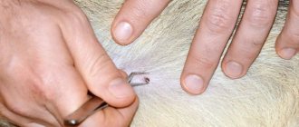
The easiest way to remove a tick is with a thread: make a loop at the end, throw it over the tick close to the skin, and tighten it. Then you need to twist the thread or make smooth circular movements with it and slowly pull it towards you until the entire tick is completely removed from the skin.
If there is still a proboscis left in the wound, it must be removed with tweezers. The bite site is treated with alcohol (or vodka, cologne) and antibiotic ointment. The tick needs to be placed in a jar and taken for analysis, see a doctor when the opportunity arises. You still need to go to the hospital, since sometimes the disease is asymptomatic in the first stages, and you can become infected from the first minutes of sucking on a tick.
In any case, after a bite, you must carefully monitor your condition; if any signs of infection appear (and they sometimes resemble only a mild cold), you should immediately seek medical help and take action.
How to protect yourself from taiga tick bites
In areas where encephalitis is endemic, vaccination against this disease is recommended for both residents and visitors.
In the absence of vaccination, the best way to protect against the threat of tick bites is repellents, which are generously available on store shelves. These can be sprays, creams, gels.
Long, light-colored clothing that covers your arms and is tucked into your shoes will be a good barrier for a tick that gets onto a person. In addition, on light-colored fabric, the bloodsucker, even if it is small from hunger, will become noticeable before it has time to attach itself.
How to protect yourself from forest ticks?
As soon as you are going to the forest, think carefully about your wardrobe
Choose suitable clothing for this purpose, which will primarily protect you. If you are bitten by a forest tick, be careful when removing it and do everything in accordance with the recommendations.
Video: how to remove forest ticks with thread
The most important thing is to be careful and attentive when traveling in areas where there is predominantly dense foliage. If you follow all preventive measures and regularly examine your own body for tick bites, you will be beyond the reach of dangerous diseases.
Video: who eats forest ticks
Preventive actions
The main tool for preventing tick-borne diseases is vaccination. During the procedure, a serum with stable resistance to viruses is injected into the person’s bloodstream, which allows the immune system to successfully cope with attacks from pathogenic microorganisms in the future. As for measures aimed at preventing tick bites, they must be comprehensive in order to completely avoid an unpleasant encounter with a pest.
Cloth
When going for a walk in the forest or park where there is a lot of greenery, you need to dress appropriately. Such measures are most relevant during periods of increased parasite activity - May, June, August, September. It is necessary to wear clothing that completely covers the body and limbs, for example:
- floor-length pants, which are better tucked into socks (in hot periods, you can wear linen clothes, they will protect the body from tick bites and be well blown by the wind, without causing discomfort during the heat);
- sneakers with socks;
- shirt with long sleeves;
- a hat with a brim (sometimes ticks climb onto the lower branches of trees, from which they can fall on the head, crawl onto the neck and start eating, so a hat with a brim will help protect against attacks from above).
When going for a walk in places where there is a high risk of being bitten by a tick, you need to dress correctly - wear long sleeves and tuck your pants into your shoes.
When walking in potentially dangerous places, it is necessary to inspect each other's clothing from time to time, paying attention to folds on the sleeves and collar. If you find an unexpected guest, you need to carefully shake him off your clothes. Taiga “villains” do not bite immediately; they can take up to one hour after contact with the skin to find the optimal place to bite.
Chemical remedies
Store-bought drugs used to combat and repel encephalitis ticks can be divided into three groups:
- repellent. These are products of various release forms containing an active substance (usually diethyltoluamide), which repels pests. The compositions are applied to clothing and exposed skin, which provides active protection against bites. For adults, drugs such as Biban, DEFI-Taiga, Gall-RET, Reftamide maximum can be used. For children, it is recommended to use less toxic products, for example, Medilisik (for children), Fthalar, Pikhtal;
- acaricidal. These are more toxic drugs made on the basis of an active component that has a nerve-paralytic effect on the tick and leads to the death of the parasitic organism (for example, alphamethrin). This group of products cannot be sprayed on the body; they are applied only to clothing, which should be spread on the ground, processed, allowed to dry, and only then put on. The most popular products are: Gardex-anti-mite, Reftamide taiga, Fumitox-anti-mite; Gardex anti-tick spray provides protection against parasites for 15 days, provided that the treated clothing is stored in a closed container when not worn.
- insecticidal and repellent. These are drugs containing both alphamethrin and diethyltoluamide in their composition, which allows them to have a lethal and repellent effect on all blood-sucking parasites, including mosquitoes, nits, and ticks. These products also cannot be applied to the skin; they are sprayed on clothes. The most popular drugs in this group are Mite-kaput aerosol, Kra-rep, Moskitol-spray.
Folk remedies
There are a lot of folk recipes and tips designed to repel ticks and prevent bites:
- Essential oil is an effective and safe preventative. To prepare the protective composition, you need to mix 2 tablespoons of olive oil with 15 drops of geranium ether, pour the resulting composition into a bottle and shake well. The product is applied to exposed skin and clothing. The drug is highly effective, but does not guarantee 100% results;
- vanillin solution. For a glass of warm water you will need 2 bags of pure vanillin - you need to carefully dilute the powder in the liquid and pour the resulting mixture into a spray bottle. The product is sprayed onto clothes and body;
- garlic. Ticks cannot tolerate the rich garlic aroma, so consuming the product will be an excellent way to prevent bites (it is recommended to take sandwiches rubbed with garlic into the forest and eat them periodically).
Pet handling
Blood-sucking creatures often attack domestic animals, which poses a danger not only to the life of the pet, but also to all household members. In order to prevent bites, it is necessary to treat with special acaricidal compounds (Advantix drops, Fiprist spray, Bolfo, etc.). They are usually applied every 3 months throughout the entire period of parasite activity. An important point is that you need to treat not only the animal, but also its place, the bedding where it sleeps, since ticks can hide reliably.
A taiga tick bite is not only unpleasant, but also potentially dangerous. These arachnids can carry and transmit severe infectious diseases through their bites, including encephalitis and Lyme disease. To minimize the risk of becoming food for the parasite, it is worth implementing a whole range of preventive measures.
Sources
- https://bez-klopa.ru/entsiklopediya/taezhnyiy-kleshh-foto-stroenie-zhiznennyiy-tsikl-chem-opasen/
- https://Dezoff.ru/kleshhej/tayezhnyy-kleshch/
- https://TaraKlop.ru/kleshchi/taezhnyj-klesch/
- https://VrediteliSOS.ru/nasekomye/kleshhi/taezhnyj-kleshch.html
- https://parazitdoma.ru/drugie-parazity/tayozhnyy-klesch
- https://givotniymir.ru/taezhnyj-kleshh-obraz-zhizni-i-sreda-obitaniya-tayozhnogo-kleshha/
- https://kleshun.ru/taezhnyj-kleshh/
- https://beetlestop.ru/taezhnyiy-kleshh/
- https://zoolog.guru/drugaya-poleznaya-informacia/taezhnyiy-kleshh-opasen-tem-chto-on-yavlyaetsya.html
- https://bugattack.ru/kleshhi/taezhnyj-kleshh.html
- https://KlopSOS.ru/kleshhi/taezhnyj-kleshh/
- https://domovod.guru/borba-s-vreditelyami/kleshhi/chem-opasen-taezhnyiy-kleshh.html
[collapse]
